Copyright
©The Author(s) 2025.
World J Nephrol. Jun 25, 2025; 14(2): 105374
Published online Jun 25, 2025. doi: 10.5527/wjn.v14.i2.105374
Published online Jun 25, 2025. doi: 10.5527/wjn.v14.i2.105374
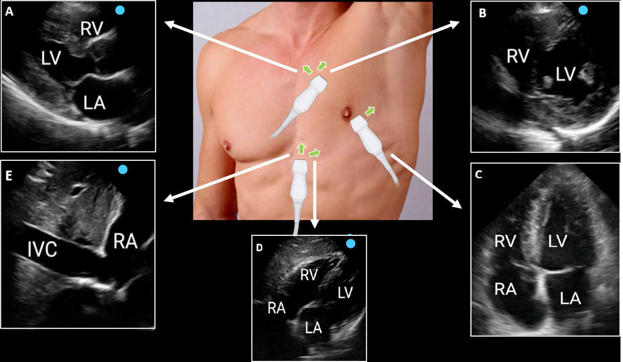
Figure 3 Focused cardiac ultrasound views.
A: Parasternal long axis; B: parasternal short axis; C: Apical four-chamber; D: Subxiphoid; E: Inferior vena cava. Green arrows indicate the direction of the transducer orientation marker. Reproduced from Argaiz et al[59]. IVC: Inferior vena cava; LA: Left atrium; LV: Left ventricle; RA: Right atrium; RV: Right ventricle. Citation: Argaiz ER, Koratala A, Reisinger N. Comprehensive Assessment of Fluid Status by Point-of-Care Ultrasonography. Kidney360 2021; 2: 1326-1338. Copyright © 2021 by the American Society of Nephrology. Published by Wolters Kluwer Health, Inc. The authors have obtained the permission for figure using (Supplementary material).
- Citation: Diniz H, Ferreira F, Koratala A. Point-of-care ultrasonography in nephrology: Growing applications, misconceptions and future outlook. World J Nephrol 2025; 14(2): 105374
- URL: https://www.wjgnet.com/2220-6124/full/v14/i2/105374.htm
- DOI: https://dx.doi.org/10.5527/wjn.v14.i2.105374


