Copyright
©The Author(s) 2016.
World J Transplant. Sep 24, 2016; 6(3): 505-516
Published online Sep 24, 2016. doi: 10.5500/wjt.v6.i3.505
Published online Sep 24, 2016. doi: 10.5500/wjt.v6.i3.505
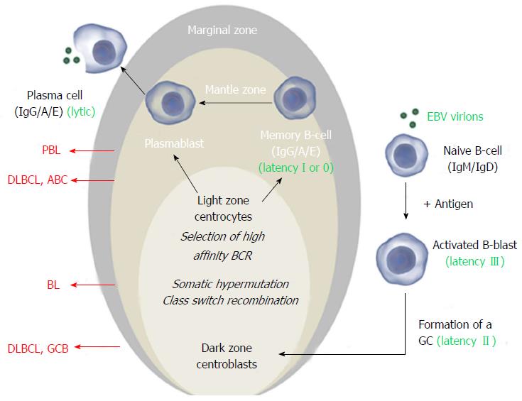
Figure 1 The Epstein-Barr virus exploits normal B-cell activation pathways.
Activation of a naive B-cell (that expresses IgM and IgD on its surface) by its cognate antigen results in B-cell activation and differentiation into a memory B-cell or a plasma cell, most commonly via T-cell dependent activation. The antigen-activated B-cell enters a primary follicle in lymph node or spleen and forms a germinal center (GC), transforming the primary follicle into a secondary follicle. This structure is composed of three distinct regions. The marginal zone[1], which consists mainly of activated B-cells and GC-matured IgM+ B-cells, the mantle zone or corona[2], which comprises naïve and memory B-cells surrounds the GC[3]. The GC consists of a dark zone and a light zone. In the dark zone, the activated B-cells (centroblasts) proliferate and downregulate expression of IgM and IgD to allow somatic hypermutation (SHM) and class switch recombination (CSR), increasing the antibody’s affinity, specificity and functional versatility. In the light zone of the GC, the B-cells (centrocytes) with the best antibody are selected and ultimately mature into memory B-cells or plasma cells. Instead of IgM and IgD, these express high affinity IgG, IgA or IgE antibodies. Classically, Epstein-Barr virus (EBV) infects naïve B-cells that are stimulated to form a GC. In the activated blast, viral latency III (LMP1+/EBNA2+) is expressed and induces proliferation. In the GC, latency II (LMP1+/EBNA2-) is expressed and infected centroblasts presumably undergo SHM and CSR, involved in antibody maturation. After leaving the GC, they differentiate into plasma cells or (mainly) memory cells (latency I, EBNA1+ or latency 0, no expression of viral proteins). In vitro and in vivo, plasma cell differentiation results in activation of the EBV lytic cycle. In all stages, the viral DNA (circle in the nucleus) is maintained as an episome. Different stages of this process can give rise to malignancy resulting in different lymphoma subtypes that have features of their normal counterpart. Here the stages at which EBV+ and EBV- B-cell lymphoma may arise are shown for the most common subtypes. Images from http://www.somersault1824.com were used in this figure. PBL: Plasmablastic lymphoma; DLBCL: Diffuse large B-cell lymphoma; ABC: Activated B-cell; GCB: Germinal center B-cell.
- Citation: Morscio J, Tousseyn T. Recent insights in the pathogenesis of post-transplantation lymphoproliferative disorders. World J Transplant 2016; 6(3): 505-516
- URL: https://www.wjgnet.com/2220-3230/full/v6/i3/505.htm
- DOI: https://dx.doi.org/10.5500/wjt.v6.i3.505


