Copyright
©The Author(s) 2016.
World J Transplant. Mar 24, 2016; 6(1): 69-90
Published online Mar 24, 2016. doi: 10.5500/wjt.v6.i1.69
Published online Mar 24, 2016. doi: 10.5500/wjt.v6.i1.69
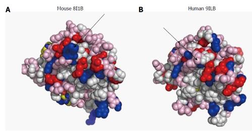
Figure 1 Surface design of mouse (A) and human (B) interleukin-1β.
The proteins are imaged at identical angles. Blue: Positively-charged amino acids; red: Negatively-charged amino acids; pink: Polar amino acids (slightly negative at physiological pH). The arrows point to differences in surface charges between the 2 proteins. Image resolved using ASAview[214].
- Citation: Barkai U, Rotem A, de Vos P. Survival of encapsulated islets: More than a membrane story. World J Transplant 2016; 6(1): 69-90
- URL: https://www.wjgnet.com/2220-3230/full/v6/i1/69.htm
- DOI: https://dx.doi.org/10.5500/wjt.v6.i1.69


