Copyright
©The Author(s) 2016.
World J Psychiatr. Mar 22, 2016; 6(1): 84-101
Published online Mar 22, 2016. doi: 10.5498/wjp.v6.i1.84
Published online Mar 22, 2016. doi: 10.5498/wjp.v6.i1.84
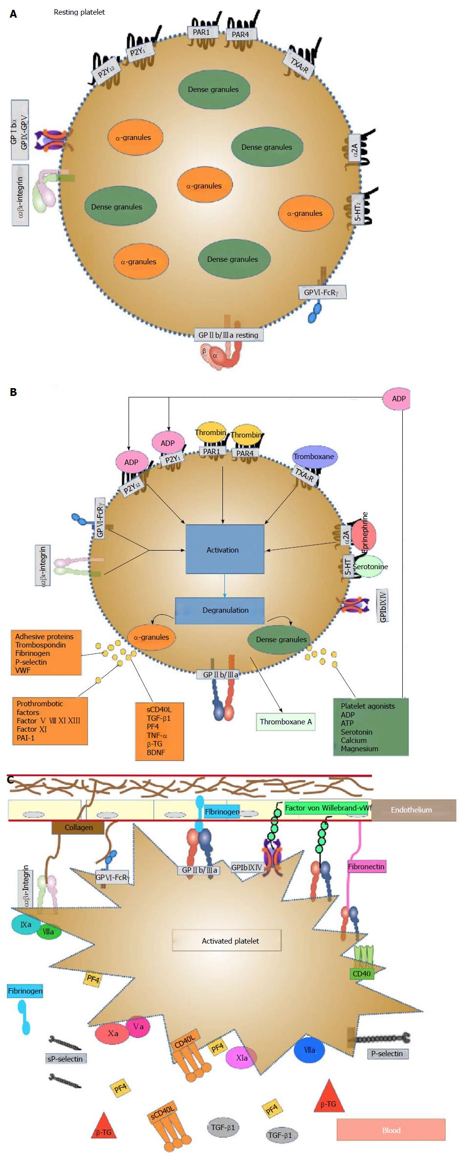
Figure 1 Representation of platelets before and after activation.
A: Resting platelet: The discoid form of resting platelets is supported by a circumferential coil of microtubules lying just below the surface membrane whereas α-granules and dense bodies are irregularly dispersed in the cytoplasm. Platelet membrane contains receptors for ADP, thrombin, vWF, fibrin, fibronectin, epinephrine, PAF, thromboxane A2, Prostacyclin, trombospondin, serotonin and gycosil transferase; B: Platelet activation: Platelets may be activated via different agonists, through several receptor-mediator pathways (thrombin, TXA2, collagen, epinephrine, etc.). Collagen exposed within the denuded area of vascular endothelium stimulates platelet activation and adhesion to the vessel wall. These agonists bind and activate their respective receptors, which in turn stimulate the activation of associated G-proteins, ultimately activating GP IIb/IIIa receptors and promoting the interaction of adjacent platelets within the clot. When the platelet is stimulated a transient influx of calcium occurs, followed by extrusion of platelet storage granule contents. The dense bodies contain serotonin (5-HT), ADP, ATP and calcium, whereas the α-granules contain the highest amount of proteins, including vWF, fibronectin, fibrinogen, P-selectin, PF-4, β-TG, RANTES, CD40, CD40L, PDGF, amyloid precursor protein, MMP, adhesion molecules (ICAM, VCAM and PECAM), various coagulation and growth factors (such as BDNF) and inflammatory markers. All these phenomena are calcium (Ca2+)-dependent, amplify platelet activation and induce irreversible platelet-platelet aggregation and thrombus formation; C: Platelet activation-aggregation: Platelet aggregation following platelet activation is marked by a shape change that increases their surface area available for adhesion and gives platelets the ability to bind fibrinogen and vWF via the active form of the surface glycoprotein GPIIb/IIIa receptors, leading to platelet aggregation and thrombus formation. Platelets express certain surface markers such as P-selectin and CD40 ligand and these expressed activation markers are cleaved, promoting the circulation of soluble CD40L and soluble P-selectin. Platelets also expose phosphatidylserine providing attachment sites for coagulation factors. Vascular injury also exposes subendothelial tissue factor, which forms a complex with factor VIIa and sets off a chain of events that culminates in the formation of the prothrombinase complex. Prothrombin is converted to thrombin, which subsequently converts fibrinogen to fibrin, generating a fibrin-rich clot. The coagulation cascade contributes to the stabilization of the thrombus. BDNF: Brain-derived neurotrophic factor; ADP: Adenosine-diphosphate; GP: Glycoprotein; ATP: Adenosine-triphosphate; PF-4: Platelet factor-4; β-TG: β-thromboglobulin; PDGF: Platelet-derived growth factor; vWF: Von Willebrand factor; MMP: Matrix metalloproteinases.
- Citation: Serra-Millàs M. Are the changes in the peripheral brain-derived neurotrophic factor levels due to platelet activation? World J Psychiatr 2016; 6(1): 84-101
- URL: https://www.wjgnet.com/2220-3206/full/v6/i1/84.htm
- DOI: https://dx.doi.org/10.5498/wjp.v6.i1.84


