Copyright
©The Author(s) 2016.
World J Psychiatr. Mar 22, 2016; 6(1): 18-30
Published online Mar 22, 2016. doi: 10.5498/wjp.v6.i1.18
Published online Mar 22, 2016. doi: 10.5498/wjp.v6.i1.18
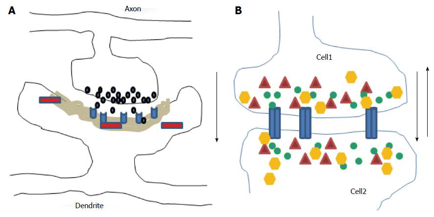
Figure 1 Diagrams illustrate chemical (A) and electrical (B) synapses.
At chemical synapses, neurotransmitters (black) released from axonal boutons bind to postsynaptic receptors (light blue) and trigger specific signaling pathways via activation of proteins (red) in postsynaptic cells with prominent postsynaptic densities component (grey area). The information transmission is unidirectional (black arrow). At electrical synapses, gap junction channels (blue) directly connect the two adjacent cells, thus enable the bidirectional passage of electrical currents (black arrows) carried by ions (green), and of small peptides (yellow) or second messengers (dark red).
- Citation: Quach TT, Lerch JK, Honnorat J, Khanna R, Duchemin AM. Neuronal networks in mental diseases and neuropathic pain: Beyond brain derived neurotrophic factor and collapsin response mediator proteins. World J Psychiatr 2016; 6(1): 18-30
- URL: https://www.wjgnet.com/2220-3206/full/v6/i1/18.htm
- DOI: https://dx.doi.org/10.5498/wjp.v6.i1.18


