Copyright
©The Author(s) 2015.
World J Hypertens. May 23, 2015; 5(2): 74-78
Published online May 23, 2015. doi: 10.5494/wjh.v5.i2.74
Published online May 23, 2015. doi: 10.5494/wjh.v5.i2.74
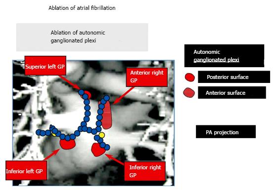
Figure 1 An anatomic figure of the left atrium depicting the lines of radiofrequency applications used to isolate the pulmonary veins in patients undergoing catheter ablation for atrial fibrillation.
Note that the lesion lines may, in part, also ablate the ganglionated plexi (GP), albeit incompletely. These nerve clusters are situated at the pulmonary vein - atrial junctions and contribute to the neural basis of atrial fibrillation[12].
- Citation: Scherlag BJ, Po SS. Symplicity-3 hypertension trial: Basic and clinical insights. World J Hypertens 2015; 5(2): 74-78
- URL: https://www.wjgnet.com/2220-3168/full/v5/i2/74.htm
- DOI: https://dx.doi.org/10.5494/wjh.v5.i2.74


