Copyright
©2012 Baishideng.
World J Exp Med. Aug 20, 2012; 2(4): 78-85
Published online Aug 20, 2012. doi: 10.5493/wjem.v2.i4.78
Published online Aug 20, 2012. doi: 10.5493/wjem.v2.i4.78
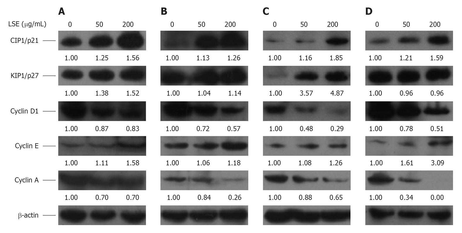
Figure 3 Immunoblots of cell cycle-controlling proteins in longan seed extract-treated cancer cells.
About 50 and 200 μg/mL longan seed extract (LSE)-treated (A) A-549, (B) Hep G2, (C) C-33A and (D) MDA-MB-231 cells were incubated at 37 °C for 48 h. The harvested cells were lysed in Triton X 100-containing hypotonic buffer as per previous reports (Hsu et al[37], 2010; Chung et al[48], 2010) at 4 °C for 30 min. Cell lysates were centrifuged and the protein concentrations in the supernatants were determined using a bicinchoninic acid protein detection kit. Cell protein lysates were separated by sodium dodecyl sulfate polyacrylamide gel electrophoresis, transferred to polyvinylidene difluoride membranes and immunoblotted to show cyclin D1, cyclin E, cyclin A, CIP1/p21 and KIP1/p27, with the β-actin level used as the loading control. The images are representative results from three independent experiments. The density of each protein band was measured using ImageJ software and protein expression was normalized to β-actin, and the relative amount of each protein band was referenced to the untreated control.
- Citation: Lin CC, Chung YC, Hsu CP. Potential roles of longan flower and seed extracts for anti-cancer. World J Exp Med 2012; 2(4): 78-85
- URL: https://www.wjgnet.com/2220-315X/full/v2/i4/78.htm
- DOI: https://dx.doi.org/10.5493/wjem.v2.i4.78


