Copyright
©The Author(s) 2023.
World J Exp Med. Sep 20, 2023; 13(4): 75-94
Published online Sep 20, 2023. doi: 10.5493/wjem.v13.i4.75
Published online Sep 20, 2023. doi: 10.5493/wjem.v13.i4.75
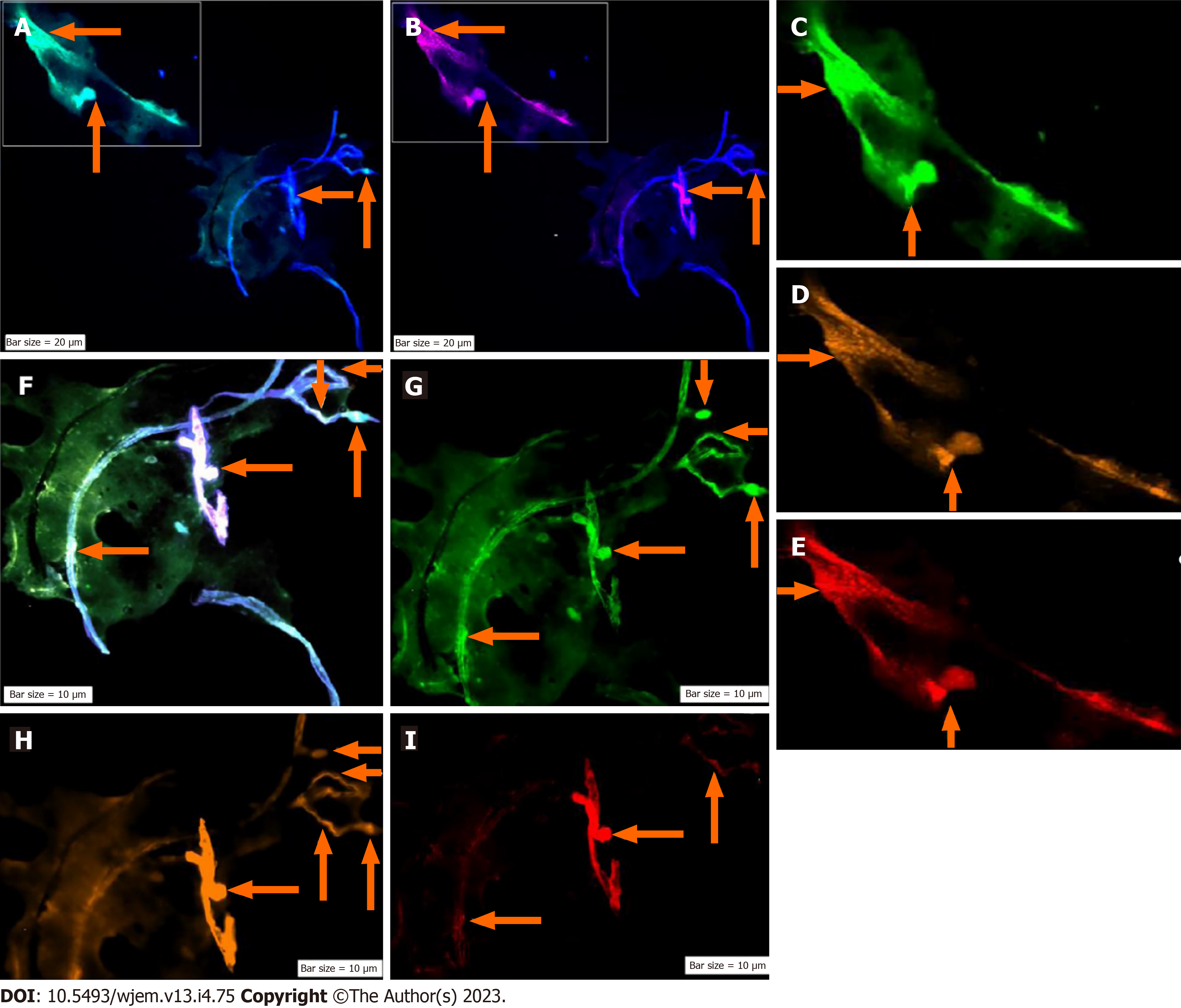
Figure 6 Protein expression of neural marker/CD133/vascular endothelial growth factor in the circulated neural cells from the neural system to the blood stream in a patient with Alzheimer disease.
A: Merged of dapi/Ne (Ne: Neural cells conjugated with dapi/Ne); B: Merged of dapi/Pe-cy5, CD133, Same cells conjugated with fluorescein isothiocyanate reflecting high expression; C: Neural cells, conjugated with fluorescein isothiocyanate (FITC)/Ne; D: Neural stem cells, conjugated with Pe-cy5 with mosaic pattern including the cells with high, very low-, and lacking-expression; E: Conjugated with Pe-cy5/neural cells; F: Co-expression of Dapi/Ne/CD133/VEGF; G: Neural cells conjugated with FITC; H: Vascular endothelial growth factor conjugated with Rpe; I: CD133 conjugated with Pe-cy5. The migrated neural cells with high or low expression are shown with arrows. The micro-vesicle and the vascular section harboring the migrated neural cells are detectable. The micro-vesicle and the vascular section harboring the migrated neural cells are detectable. Ne: Neural cells conjugated with FITC; CD133: Neural stem cells, conjugated with Pe-cy5; VEGF: Vascular endothelial growth factor, conjugated with RPe; The CNCs were explored by IF method for combination of neural marker, neural stem cell for Ne/CD133 (Figure 7).
- Citation: Mehdipour P, Fathi N, Nosratabadi M. Personalized clinical managements through exploring circulating neural cells and electroencephalography. World J Exp Med 2023; 13(4): 75-94
- URL: https://www.wjgnet.com/2220-315X/full/v13/i4/75.htm
- DOI: https://dx.doi.org/10.5493/wjem.v13.i4.75


