Copyright
©The Author(s) 2016.
World J Crit Care Med. Feb 4, 2016; 5(1): 74-82
Published online Feb 4, 2016. doi: 10.5492/wjccm.v5.i1.74
Published online Feb 4, 2016. doi: 10.5492/wjccm.v5.i1.74
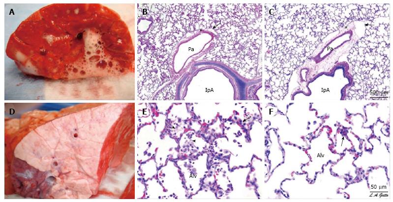
Figure 1 A gross and histological comparison between airway pressure release ventilation and nonpreventative ventilation.
A and D: Gross pathology of the cut surface of the right lower lobe of the lung of representative animals from (A) the NPV and (D) the APRV group. The NPV shows severe inflammation, bronchial edema, and areas of hemorrhage. The APRV group demonstrates normal, pink, homogenously inflated lungs with little injury on gross appearance; B, C, E, F: Histological comparison of four pigs, two NPV (B and E) and two APRV (C and F) at low (B and C) and high (E and F) magnification. The NPV animals show classic stigmata of ARDS including atelectasis, fibrinous exudates, intra-alveolar hemorrhage, congested capillaries, thickened alveolar walls, and leukocytic infiltrates. The APRV animals demonstrate preservation of nearly normal pulmonary architecture. Published with permssion from Ref[38]. APRV: Airway pressure release ventilation; NPV: Nonpreventative ventilation; ARDS: Acute respiratory distress syndrome; Alv: Alveoli.
- Citation: Sadowitz B, Jain S, Kollisch-Singule M, Satalin J, Andrews P, Habashi N, Gatto LA, Nieman G. Preemptive mechanical ventilation can block progressive acute lung injury. World J Crit Care Med 2016; 5(1): 74-82
- URL: https://www.wjgnet.com/2220-3141/full/v5/i1/74.htm
- DOI: https://dx.doi.org/10.5492/wjccm.v5.i1.74


