Copyright
©The Author(s) 2016.
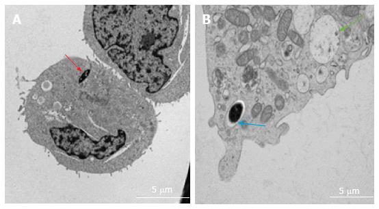
Figure 9 Transmission electron microscopy images of intracellular localisation of mycobacteria in human mast cell line (A) and bone marrow derived mast cells (B).
Cells infected with Mycobacterium marinum at MOI 10 for 24 h. A, red arrow: Cytoplasmic localisation; B, blue arrow: Vacuolar localisation; green arrow: Empty vacuole comparable to those described for degranulating mast cells[11]. MOI: Multiplicity of infection.
- Citation: Siad S, Byrne S, Mukamolova G, Stover C. Intracellular localisation of Mycobacterium marinum in mast cells. World J Immunol 2016; 6(1): 83-95
- URL: https://www.wjgnet.com/2219-2824/full/v6/i1/83.htm
- DOI: https://dx.doi.org/10.5411/wji.v6.i1.83


