Copyright
©The Author(s) 2015.
World J Respirol. Nov 28, 2015; 5(3): 188-198
Published online Nov 28, 2015. doi: 10.5320/wjr.v5.i3.188
Published online Nov 28, 2015. doi: 10.5320/wjr.v5.i3.188
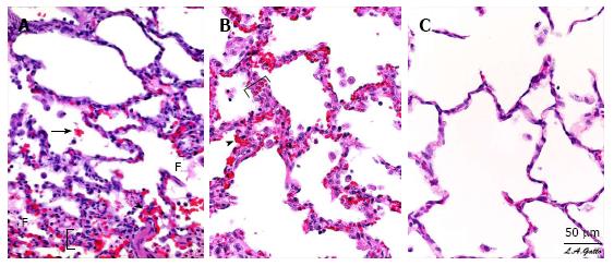
Figure 3 Pulmonary Histopathology - Photomicrographs of representative lung sections of specimens from each treatment group at 40 x magnification.
A: Sham- animals received 48 h of mechanical ventilation without peritoneal sepsis + gut ischemia/reperfusion (PS + I/R) injury. Specimen exhibits stigmata of lung injury including fibrinous deposits, blood in alveolus, congested capillaries, and thickened alveolar walls; B: LTV- animals received PS + I/R injury and LTV ventilation after onset of ALI. Specimen exhibits stigmata of lung injury including fibrinous deposits, blood in alveolus, congested capillaries, leukocyte infiltration, and thickened alveolar walls; C: APRV- Animals received APRV one hour following PS + I/R injury. Specimen shows normal pulmonary architecture, alveoli are well expanded, thin walled and there are no exudates. APRV applied early in animals with severe septic shock protected the lung superior to Sham animals with conventional mechanical ventilation without septic shock. (with permission)[33]. F: Fibrinous deposit in the air compartment; Arrow: Blood in alveolus; Arrowhead: Congested alveolar capillary; Bracket: Thickened alveolar wall; APRV: Airway pressure release ventilation.
- Citation: Nieman GF, Gatto LA, Habashi NM. Reducing acute respiratory distress syndrome occurrence using mechanical ventilation. World J Respirol 2015; 5(3): 188-198
- URL: https://www.wjgnet.com/2218-6255/full/v5/i3/188.htm
- DOI: https://dx.doi.org/10.5320/wjr.v5.i3.188


