Copyright
©The Author(s) 2016.
World J Ophthalmol. May 12, 2016; 6(2): 10-19
Published online May 12, 2016. doi: 10.5318/wjo.v6.i2.10
Published online May 12, 2016. doi: 10.5318/wjo.v6.i2.10
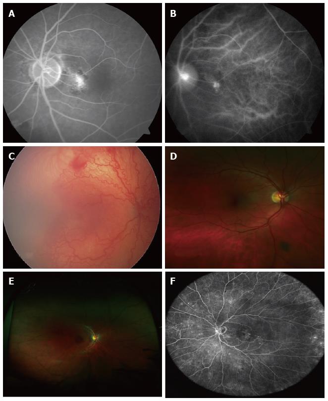
Figure 2 Advanced retinal imaging with and without contrast.
A: Fluorescein angiography. Localised hyperfluorescence; B: ICG revealing the source of leakage depicted on FFA, Figure 2A: Idiopathic polypoidal choroidal vascularisation; C: UWF fundus photo showing ROP taken with Retcam; D: Optomap ultra-widefield retinal image. Peripheral pigmented lesion not visible on standard colour fundus photo; E: Optomap ultra-widefield retinal image. Note lash artefact; F: Heidelberg HRA ultra-widefield retinal image. ICG: Indocyanine green; FFA: Fundus fluorescein angiography; UWF: Ultra-wide field; ROP: Retinopathy of prematurity.
- Citation: Saeed MU, Oleszczuk JD. Advances in retinal imaging modalities: Challenges and opportunities. World J Ophthalmol 2016; 6(2): 10-19
- URL: https://www.wjgnet.com/2218-6239/full/v6/i2/10.htm
- DOI: https://dx.doi.org/10.5318/wjo.v6.i2.10


