Copyright
©The Author(s) 2016.
World J Ophthalmol. May 12, 2016; 6(2): 10-19
Published online May 12, 2016. doi: 10.5318/wjo.v6.i2.10
Published online May 12, 2016. doi: 10.5318/wjo.v6.i2.10
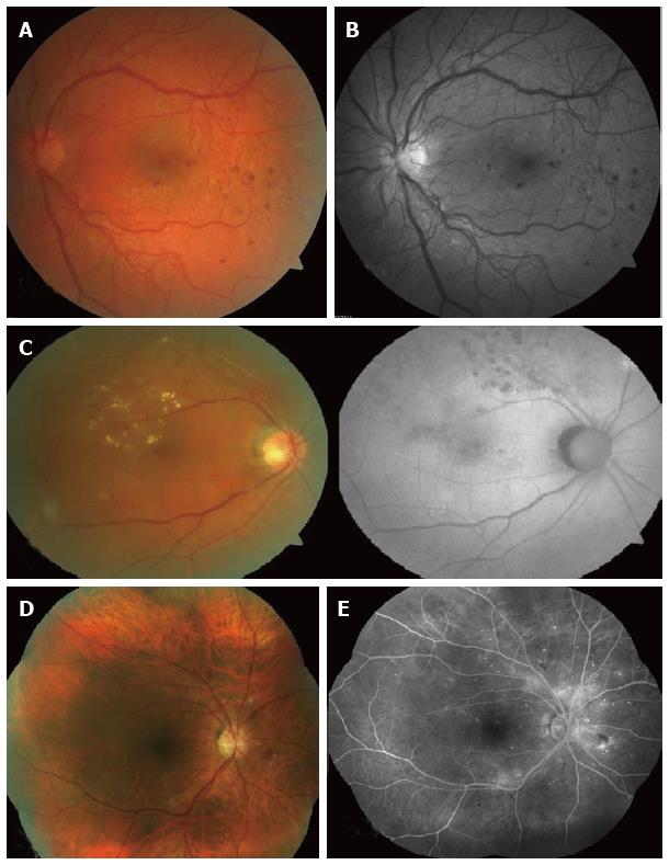
Figure 1 Retinal photographs by various funds cameras.
A: Colour fundus photo showing left eye with proliferative diabetic retinopathy; (Image taken by Zeiss Cirrus photo 800). Compare with Figure 1B; B: Red-free fundus photo (left eye with proliferative diabetic retinopathy). Better visualisation of new vessels and retinal haemorrhages as opposed to Figure 1A; C: Colour fundus photo (left) vs fundus autofluorescence (right); Retinal exudates are more visible on the colour fundus photo. Autofluorescence highlights the laser scars not readily visible on a colour fundus photo; D: Seven-field fundus photo - computer-aided mosaic (Image taken by Zeiss Cirrus 800); E: Seven-field fundus with fluorescein angiography (Image taken by Zeiss Visucam D500).
- Citation: Saeed MU, Oleszczuk JD. Advances in retinal imaging modalities: Challenges and opportunities. World J Ophthalmol 2016; 6(2): 10-19
- URL: https://www.wjgnet.com/2218-6239/full/v6/i2/10.htm
- DOI: https://dx.doi.org/10.5318/wjo.v6.i2.10


