Copyright
©The Author(s) 2015.
World J Neurol. Dec 28, 2015; 5(4): 113-131
Published online Dec 28, 2015. doi: 10.5316/wjn.v5.i4.113
Published online Dec 28, 2015. doi: 10.5316/wjn.v5.i4.113
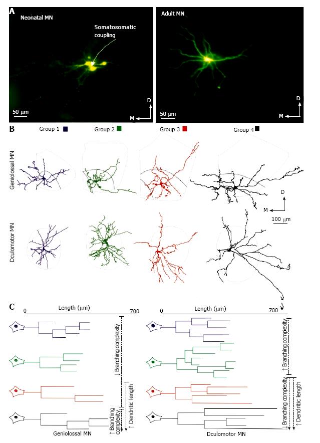
Figure 2 Summary of the main morphological changes of motoneurons from genioglossal and oculomotor nuclei.
A: Photomicrographs of representative GG MNs injected intracellularly with Lucifer yellow in a newborn (left image) and juvenile adult rat (right image). Note the presence of coupling between neonatal MNs. Only one cell was injected, however 3-4 are labeled; B: Changes in nucleus size and dendritic arborization orientation of MNs during postnatal development. It is remarkable that dendrites in the younger MNs are restricted to the nucleus boundaires, whereas the 2 oldest groups show some portions of dendrites outside those limits, preferably in the ventrolateral axis. The outer line indicates the boundary of the nuclei, and the inner line shows the limits of the GG nucleus; C: Dendrograms for a representative dendrite for each age group of GG (left) and OCM (right) MNs. Note the growth in length for both groups of MNs and the changes in dendrite architecture that take place during development. The data from GG[3,5] and OCM[11] come from previous studies. MNs: Motoneurons; GG: Genioglossal; OCM: Oculomotor.
- Citation: Carrascal L, Nieto-González J, Pardillo-Díaz R, Pásaro R, Barrionuevo G, Torres B, Cameron WE, Núñez-Abades P. Time windows for postnatal changes in morphology and membrane excitability of genioglossal and oculomotor motoneurons. World J Neurol 2015; 5(4): 113-131
- URL: https://www.wjgnet.com/2218-6212/full/v5/i4/113.htm
- DOI: https://dx.doi.org/10.5316/wjn.v5.i4.113


