Copyright
©The Author(s) 2015.
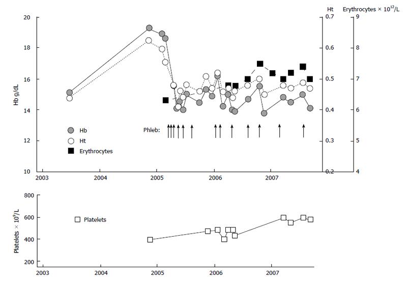
Figure 13 Clinical course in case 8 (Table 9, Figure 11) with erythrocythemic polycythemia vera treated with venesections (arrows).
The development of microcytic hypochromic erythrocytes due to iron deficiency was associated with persistent increased red cell count (> 6 × 10¹²/L), which is diagnostic for polycythemia vera. The iron deficient state and the low normal values for haemoglobin (Hb) and hematocrit (Ht) was associated with relief of hypervolumic symptoms with phlebotomy on top of low dose aspirin.
-
Citation: Michiels JJ, Valster F, Wielenga J, Schelfout K, Raeve HD. European
vs 2015-World Health Organization clinical molecular and pathological classification of myeloproliferative neoplasms. World J Hematol 2015; 4(3): 16-53 - URL: https://www.wjgnet.com/2218-6204/full/v4/i3/16.htm
- DOI: https://dx.doi.org/10.5315/wjh.v4.i3.16


