Copyright
©2014 Baishideng Publishing Group Co.
World J Anesthesiol. Mar 27, 2014; 3(1): 96-104
Published online Mar 27, 2014. doi: 10.5313/wja.v3.i1.96
Published online Mar 27, 2014. doi: 10.5313/wja.v3.i1.96
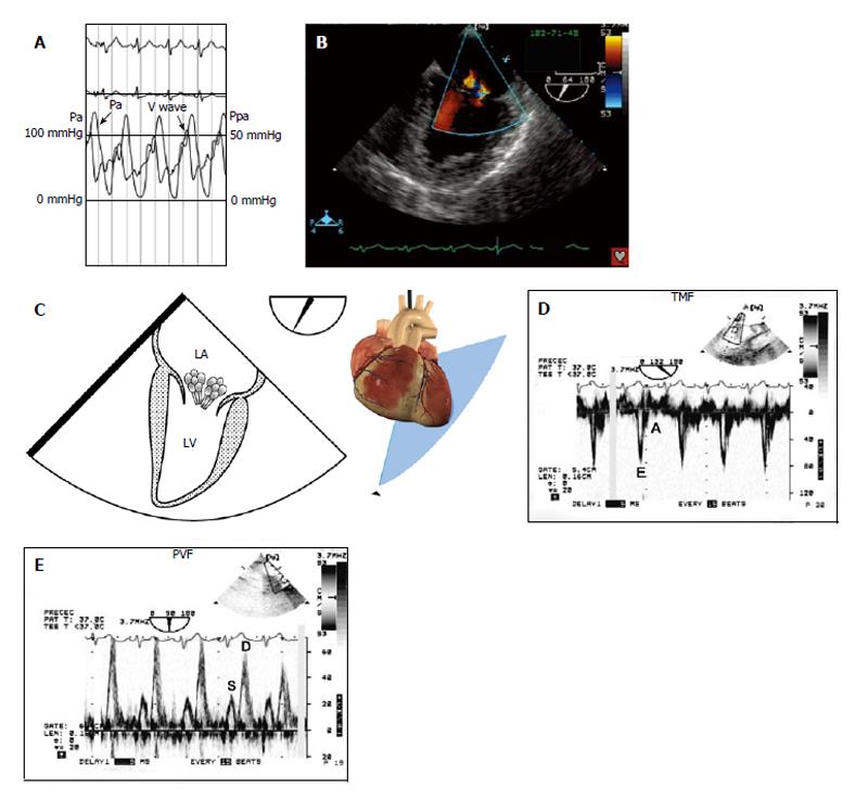
Figure 3 Stage III left ventricle diastolic dysfunction (restrictive filling).
A 61-year-old woman with cardiogenic shock is brought to the operating room for emergency coronary revascularization. A: She was hemodynamically unstable on an intra-aortic balloon pump and vasoactive support; B and C: A 50 mmHg “V” wave on the wedged pulmonary artery catheter tracing was seen without any significant mitral regurgitation on color flow imaging; D: TMF showed an E/A ratio > 2 with a deceleration time < 60 ms and isovolumic relaxation time of 40 ms; E: The left upper PVF showed an abnormal S/D ratio with S wave blunting; A: Peak late diastolic TMF velocity; D: Peak diastolic PVF velocity; E: Peak early diastolic TMF velocity. LA: Left atrium; LV: Left ventricle; Pa: Arterial pressure; Ppa: Pulmonary arterial pressure; PVF: Pulmonary venous flow; S: Peak systolic PVF velocity; TMF: Transmitral flow. With permission of Informa Healthcare adapt from reference[36].
- Citation: Denault AY, Couture P. Practical diastology. World J Anesthesiol 2014; 3(1): 96-104
- URL: https://www.wjgnet.com/2218-6182/full/v3/i1/96.htm
- DOI: https://dx.doi.org/10.5313/wja.v3.i1.96


