Copyright
©2014 Baishideng Publishing Group Co.
World J Anesthesiol. Mar 27, 2014; 3(1): 96-104
Published online Mar 27, 2014. doi: 10.5313/wja.v3.i1.96
Published online Mar 27, 2014. doi: 10.5313/wja.v3.i1.96
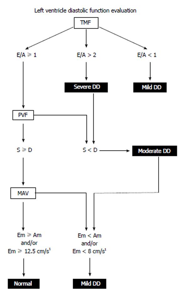
Figure 2 Left ventricle diastolic dysfunction algorithm.
LV diastolic function is classified using pulsed wave Doppler of the TMF, PVF and tissue Doppler examination of MAV. Patients with a pacemaker, atrial fibrillation, non-sinus rhythm, mitral stenosis, severe mitral and aortic regurgitation are excluded from analysis. 1Normal Em is within an 8-12.5 cm/s interval. A: Peak late diastolic TMF velocity; Am: Peak late diastolic MAV; D: Peak diastolic PVF velocity; DD: Diastolic dysfunction; E: Peak early diastolic TMF velocity; Em: Peak early diastolic MAV; LV: Left ventricle; MAV: Mitral annular velocity; PVF: Pulmonary venous flow; S: Peak systolic PVF velocity; TMF: Transmitral flow. With permission of Informa Healthcare adapt from reference[36].
- Citation: Denault AY, Couture P. Practical diastology. World J Anesthesiol 2014; 3(1): 96-104
- URL: https://www.wjgnet.com/2218-6182/full/v3/i1/96.htm
- DOI: https://dx.doi.org/10.5313/wja.v3.i1.96


