Copyright
©The Author(s) 2015.
World J Orthop. Sep 18, 2015; 6(8): 641-648
Published online Sep 18, 2015. doi: 10.5312/wjo.v6.i8.641
Published online Sep 18, 2015. doi: 10.5312/wjo.v6.i8.641
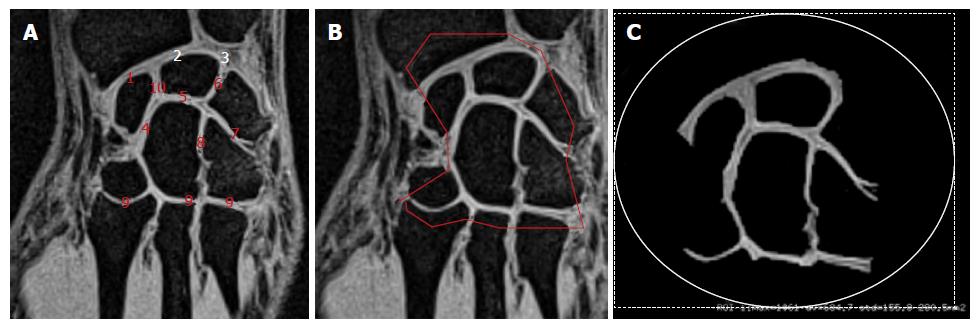
Figure 2 Manual segmentation of the cartilage cross-sectional area.
A: Coronal slice (as in Figure 1C) showing different joints between radius and scaphoid (1), radius and lunate (2), lunate and ulna (3), scaphoid and capitate (4), lunate and ulna (5), lunate and triquetrum (6), triquetrum and hamate (7) hamate and capitate (8), carpals bones and metacarpal bones (9) scaphoid and lunate (10); B: Coronal slice illustrating the manual segmentation of the cartilage area of interest and the corresponding result (C).
- Citation: Zink JV, Souteyrand P, Guis S, Chagnaud C, Fur YL, Militianu D, Mattei JP, Rozenbaum M, Rosner I, Guye M, Bernard M, Bendahan D. Standardized quantitative measurements of wrist cartilage in healthy humans using 3T magnetic resonance imaging. World J Orthop 2015; 6(8): 641-648
- URL: https://www.wjgnet.com/2218-5836/full/v6/i8/641.htm
- DOI: https://dx.doi.org/10.5312/wjo.v6.i8.641


