Copyright
©The Author(s) 2015.
World J Orthop. Sep 18, 2015; 6(8): 577-589
Published online Sep 18, 2015. doi: 10.5312/wjo.v6.i8.577
Published online Sep 18, 2015. doi: 10.5312/wjo.v6.i8.577
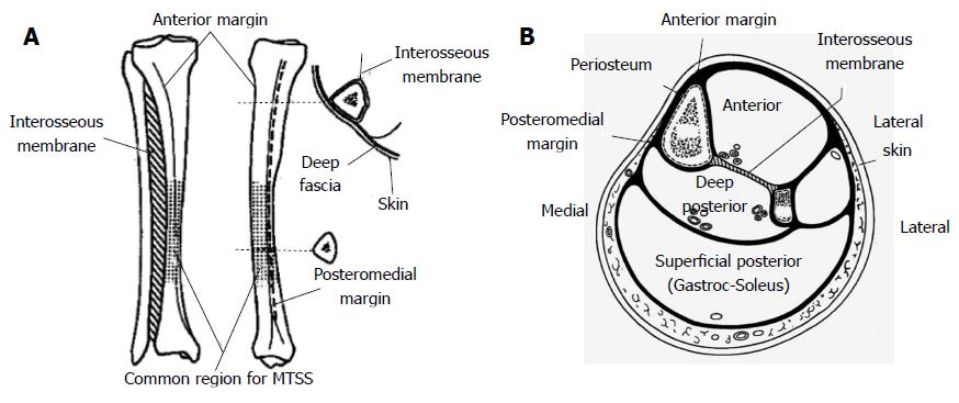
Figure 1 Anterior and medial views of the tibia with the main features shown, with the larger insert demonstrating the deep fascial attachments (A) and schematic section through the tibia illustrating the four compartments of the leg and their fascial coverings (B).
The wide subcutaneous medial surface of the tibia can be seen. Images adapted from Oakes[24].
- Citation: Franklyn M, Oakes B. Aetiology and mechanisms of injury in medial tibial stress syndrome: Current and future developments. World J Orthop 2015; 6(8): 577-589
- URL: https://www.wjgnet.com/2218-5836/full/v6/i8/577.htm
- DOI: https://dx.doi.org/10.5312/wjo.v6.i8.577


