Copyright
©2014 Baishideng Publishing Group Co.
World J Orthop. Apr 18, 2014; 5(2): 100-106
Published online Apr 18, 2014. doi: 10.5312/wjo.v5.i2.100
Published online Apr 18, 2014. doi: 10.5312/wjo.v5.i2.100
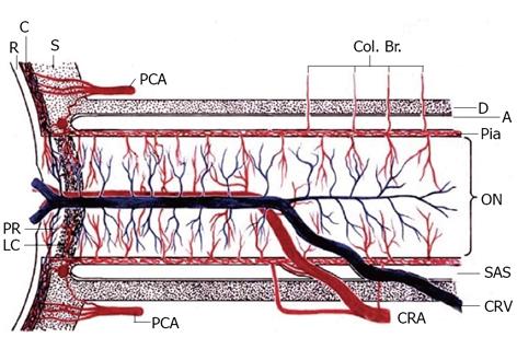
Figure 1 Schematic representation of blood supply of the optic nerve.
Reproduced from Hayreh et al[20]. A: Arachnoid; C: Choroid; CRA: Central retinal artery; Col. Br.: Collateral branches; CRV: Central retinal vein; D: Dura; LC: Lamina cribrosa; ON: Optic nerve; P: Pia; PCA: Posterior ciliary artery; PR: Prelaminar region; R: Retina, S: Sclera; SAS: Subarachnoid space.
- Citation: Nickels TJ, Manlapaz MR, Farag E. Perioperative visual loss after spine surgery. World J Orthop 2014; 5(2): 100-106
- URL: https://www.wjgnet.com/2218-5836/full/v5/i2/100.htm
- DOI: https://dx.doi.org/10.5312/wjo.v5.i2.100


