Copyright
©2013 Baishideng Publishing Group Co.
World J Orthop. Oct 18, 2013; 4(4): 241-247
Published online Oct 18, 2013. doi: 10.5312/wjo.v4.i4.241
Published online Oct 18, 2013. doi: 10.5312/wjo.v4.i4.241
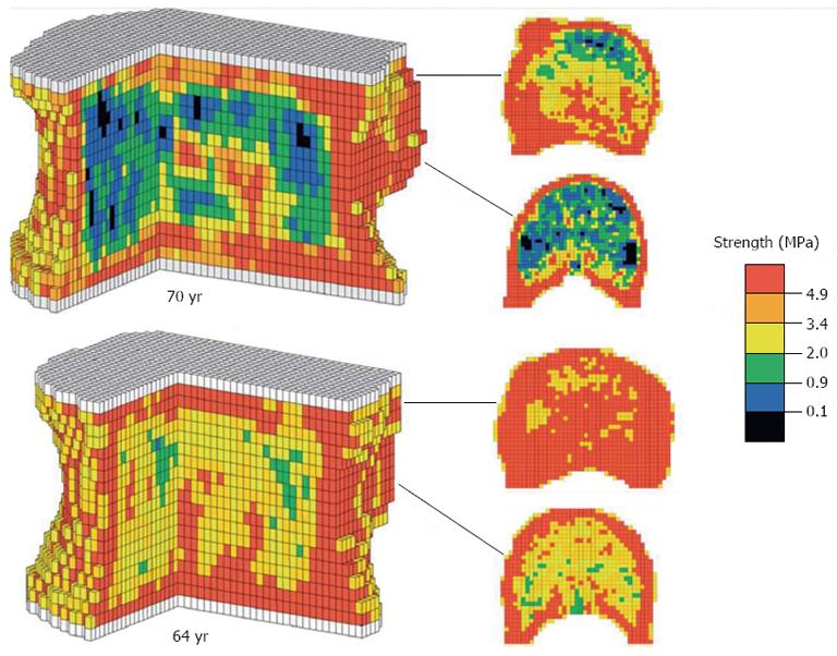
Figure 2 Three-dimensional quantitative computed tomography-based finite element modelling of the vertebral body.
The L3 vertebra from two individuals (one aged 70 and the second 64 years) modelled using voxels from 3D quantitative computed tomography, allowing estimation of bone mineral density. The model has been used to predict vertebral strength under axial compression, as illustrated by the colour scale. Overall vertebral strength were predicted as 4656 N in the younger patient (64 years) vs 6095 N in the older patient (70 years). Reproduced from Melton et al[5].
- Citation: Sisodia GB. Methods of predicting vertebral body fractures of the lumbar spine. World J Orthop 2013; 4(4): 241-247
- URL: https://www.wjgnet.com/2218-5836/full/v4/i4/241.htm
- DOI: https://dx.doi.org/10.5312/wjo.v4.i4.241


