Copyright
©The Author(s) 2023.
World J Orthop. Mar 18, 2023; 14(3): 155-165
Published online Mar 18, 2023. doi: 10.5312/wjo.v14.i3.155
Published online Mar 18, 2023. doi: 10.5312/wjo.v14.i3.155
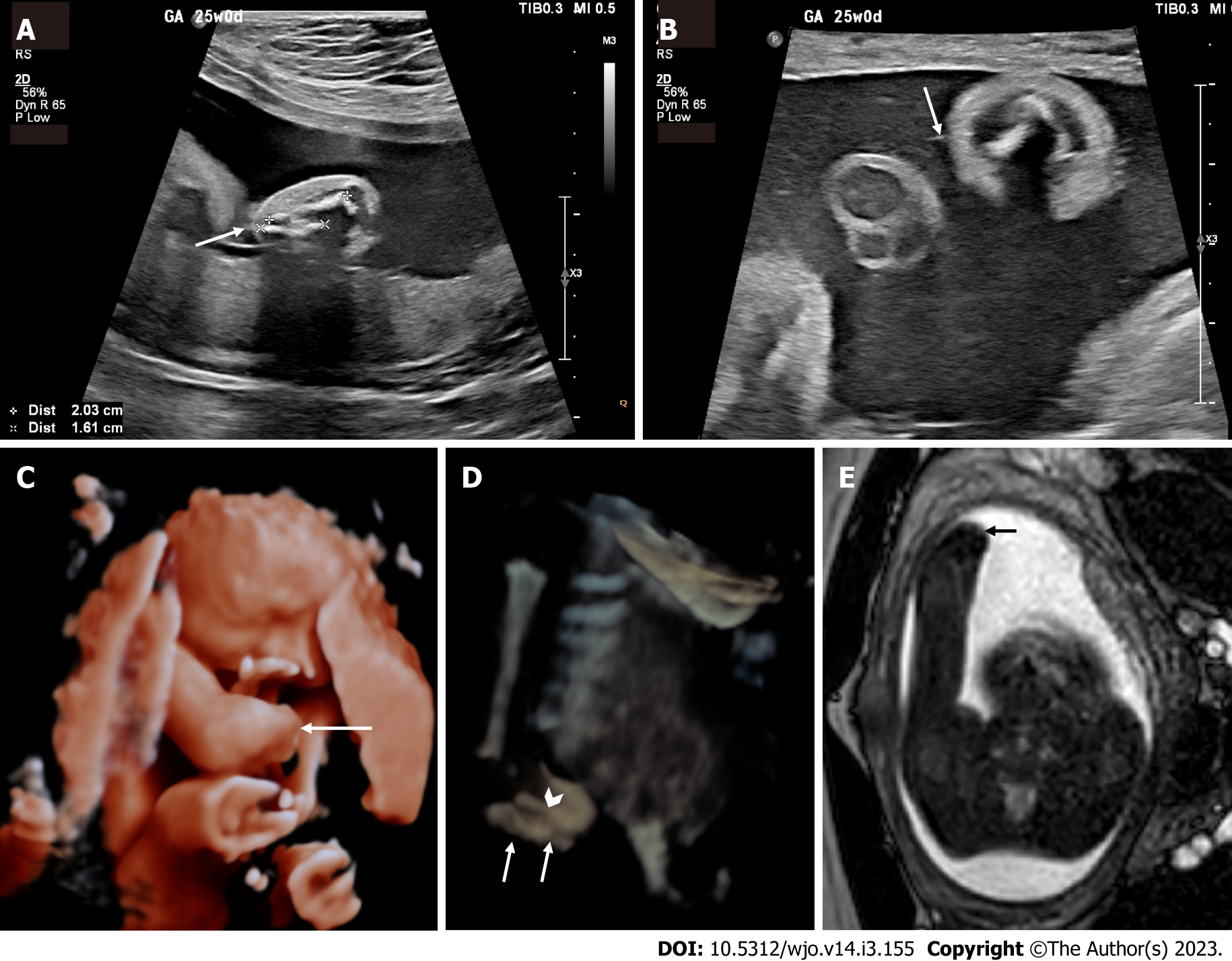
Figure 1 Prenatal imaging at 24 wk and 5 d demonstrating a transverse limb deficiency of the right forearm.
A: Two-dimensional ultrasound (2D US) showing markedly shortened right ulna (bone between "+" crosshairs) and radius (bone between "x" crosshairs) secondary to a transverse limb reduction defect (arrow); B: High-resolution 2D ultrasound with high-frequency 4-18 Megahertz probe demonstrating a hyperechogenic thin line (arrow) representing a visualized amniotic band attached to the forearm defect; C: 3D US rendered image showing the terminal transverse limb defect below the elbow (arrow); D: 3D US reconstructed image using the maximum intensity projection to demonstrate the markedly short ulna (arrows) and radius (arrowhead). The advantage of 3D US in this case is that the normal humerus and elbow (which are not in the same plane of section in Figure 1A) as well as their relationship with the amputated distal forearm can be appreciated in a single image. The 3D rendered images (Figure 1C and D) are easier to understand for both the referring providers and parents; E: Axial balanced turbo field echo fetal magnetic resonance imaging slice showing the deficiency (arrow).
- Citation: Vij N, Goncalves LF, Llanes A, Youn S, Belthur MV. Prenatal radiographic evaluation of congenital transverse limb deficiencies: A scoping review. World J Orthop 2023; 14(3): 155-165
- URL: https://www.wjgnet.com/2218-5836/full/v14/i3/155.htm
- DOI: https://dx.doi.org/10.5312/wjo.v14.i3.155


