Copyright
©The Author(s) 2024.
World J Clin Oncol. Sep 24, 2024; 15(9): 1157-1167
Published online Sep 24, 2024. doi: 10.5306/wjco.v15.i9.1157
Published online Sep 24, 2024. doi: 10.5306/wjco.v15.i9.1157
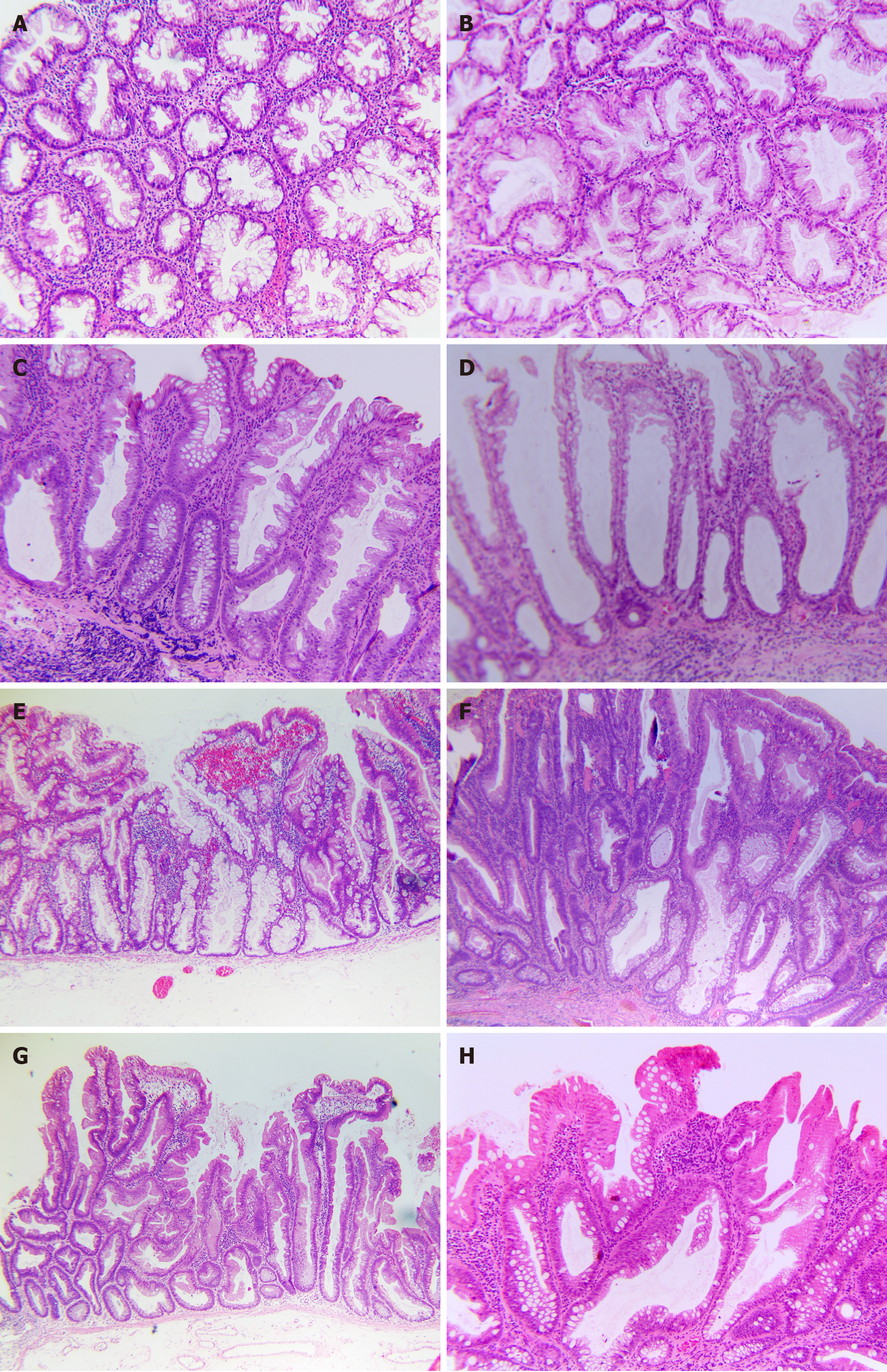
Figure 2 Chromatogram by general biopsy-specimen processing techniques.
A and B: Poorly orientated, horizontal sections (H&E, 100 ×); A: Case No. 1 with serrated pattern might require more information on polyp size and location to guide differential diagnosis between hyperplastic polyp (HP) and sessile serrated lesion (SSL); B: Case No. 2, which revealed hypercellularity and structure asymmetry, highly suggested a SSL with dysplasia (SSLD). Therefore, MLH1 staining was required for confirmative diagnosis; C and D: Crypt-base or crypt dilation might be hard to define in equivocal cases (H&E, 100 ×); C: Case No. 3 is rugged to evaluate for crypt dilation; however, the serrated feature could support diagnosis as a SSL; D: Case No. 4, which only showed the crypt base dilated without other features, could lead to being misdiagnosed or debated; E and F: Well-performed endoscopic submucosal dissection specimen with the reserved orientation of SSL and SSLD; E: Case No. 5, SSL, demonstrated the lateral growth along muscularis mucosae, creating an L or an inverted T, the most reliable feature (H&E, 40 ×); F: Case No. 6, SSLD, by World Health Organization 2019, reclassified as a mixture of serrated and intestinal dysplasia by Cenaj et al[23] and reclassified as dysplasia not specified by Liu et al[24], and reclassified as SSLDnos by American Journal of Clinical Pathology 2021 (100 ×); G and H: Several uncommon cases of serrated lesions; G: Case No. 7, which was flat overall, should be categorized as traditional serrated adenoma, transforming from an SSL rather than SSLD (H&E, 40 ×); H: Case No. 8, SSL, which had an eosinophilic surface change, should not be interpreted as cytological dysplasia (H&E, 100 ×).
- Citation: Tran TH, Nguyen VH, Vo DT. How to "pick up" colorectal serrated lesions and polyps in daily histopathology practice: From terminologies to diagnostic pitfalls. World J Clin Oncol 2024; 15(9): 1157-1167
- URL: https://www.wjgnet.com/2218-4333/full/v15/i9/1157.htm
- DOI: https://dx.doi.org/10.5306/wjco.v15.i9.1157


