Copyright
©The Author(s) 2024.
World J Clin Oncol. Jul 24, 2024; 15(7): 867-894
Published online Jul 24, 2024. doi: 10.5306/wjco.v15.i7.867
Published online Jul 24, 2024. doi: 10.5306/wjco.v15.i7.867
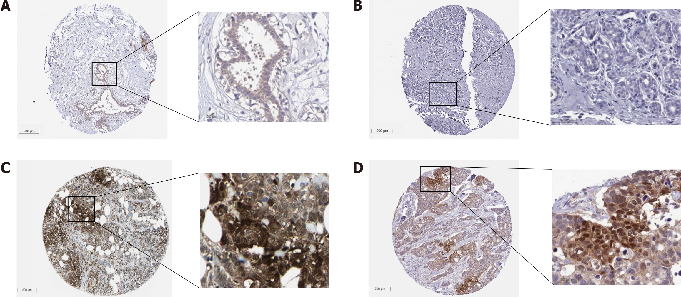
Figure 16 Immunohistochemistry staining from the Human Protein Atlas showing low or no expression of phosphoglycerate kinase 1 in normal breast tissue cells, and high expression in breast cancer cells.
A: HPA045385, female, age 45, breast (T-04000), normal tissue, NOS (M-00100), Patient ID: 3544 (low or no expression). (1) Adipocytes. Staining: Not detected; Intensity: Weak; Quantity: < 25%; Location: cytoplasmic/membranous; (2) Glandular cells. Staining: Low; Intensity: Weak; Quantity: > 75%; Location: cytoplasmic/membranous; and (3) Myoepithelial cells. Staining: Low; Intensity: Weak; Quantity: > 75%; Location: Cytoplasmic/membranous; B: HPA073644, female, age 43, breast (T-04000), skin (T-01000), normal tissue, NOS (M-00100), Patient ID: 2104 (low or no expression). Adipocytes, glandular cells, and myoepithelial cells. Staining: Not detected; Intensity: Negative; Quantity: None; C: CAB010065, female, age 59, breast (T-04000), lobular carcinoma (M-85203), Patient ID: 2805 (high expression). (1) Tumor cells. Staining: High; Intensity: Strong; Quantity: 75%-25%; Location: Cytoplasmic/membranous nuclear; D: HPA045385, female, age 83, breast (T-04000), duct carcinoma (M-85003), Patient ID: 2160 (high expression). (1) Tumor cells. Staining: High. Intensity: Strong. Quantity: 75%-25%. Location: Cytoplasmic/membranous nuclear. These immunohistochemical protein expression figures were from the Human Protein Atlas (THPA) database [71].
- Citation: Chen JY, Li JD, He RQ, Huang ZG, Chen G, Zou W. Bibliometric analysis of phosphoglycerate kinase 1 expression in breast cancer and its distinct upregulation in triple-negative breast cancer. World J Clin Oncol 2024; 15(7): 867-894
- URL: https://www.wjgnet.com/2218-4333/full/v15/i7/867.htm
- DOI: https://dx.doi.org/10.5306/wjco.v15.i7.867


