Copyright
©The Author(s) 2024.
World J Clin Oncol. Feb 24, 2024; 15(2): 178-194
Published online Feb 24, 2024. doi: 10.5306/wjco.v15.i2.178
Published online Feb 24, 2024. doi: 10.5306/wjco.v15.i2.178
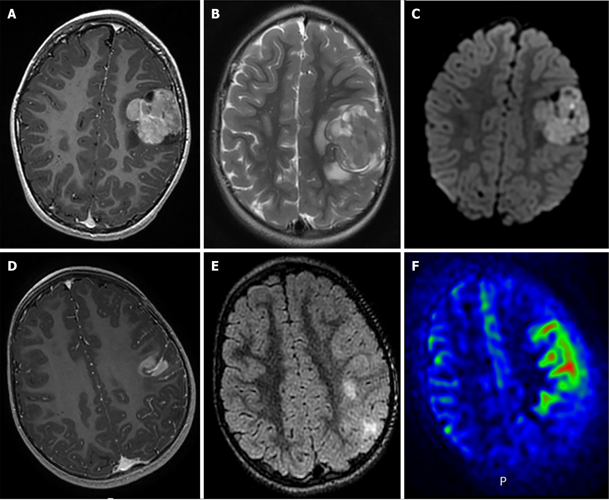
Figure 5 Radiological features of two supratentorial pediatric high-grade gliomas of the subclass MYCN.
A-C: T1-weighted images after contrast media injection (A), T2-weighted images (B), and diffusion-weighted images (C): a solid lesion with peri-lesional edema, homogeneous enhancement and hypercellularity (apparent diffusion coefficient (ADC) on diffusion weighted images is restricted in the main part of the tumor) (Case 3); D-F: T1-weighted images after contrast media injection (D), FLAIR-weighted images (E) and cerebral blood flow map using arterial spin labeling (F): a solid and infiltrative lesion with homogeneous enhancement and high cerebral blood flow (Case 1). Citation: Tauziède-Espariat A, Debily MA, Castel D, Grill J, Puget S, Roux A, Saffroy R, Pagès M, Gareton A, Chrétien F, Lechapt E, Dangouloff-Ros V, Boddaert N, Varlet P. The pediatric supratentorial MYCN-amplified high-grade gliomas methylation class presents the same radiological, histopathological and molecular features as their pontine counterparts. Acta Neuropathol Commun 2020; 8: 104. Copyright ©2020 The Authors. Published by BioMed Central Ltd[100] (Supplementary material).
- Citation: Mohamed AA, Alshaibi R, Faragalla S, Mohamed Y, Lucke-Wold B. Updates on management of gliomas in the molecular age. World J Clin Oncol 2024; 15(2): 178-194
- URL: https://www.wjgnet.com/2218-4333/full/v15/i2/178.htm
- DOI: https://dx.doi.org/10.5306/wjco.v15.i2.178


