Copyright
©The Author(s) 2024.
World J Clin Oncol. Feb 24, 2024; 15(2): 178-194
Published online Feb 24, 2024. doi: 10.5306/wjco.v15.i2.178
Published online Feb 24, 2024. doi: 10.5306/wjco.v15.i2.178
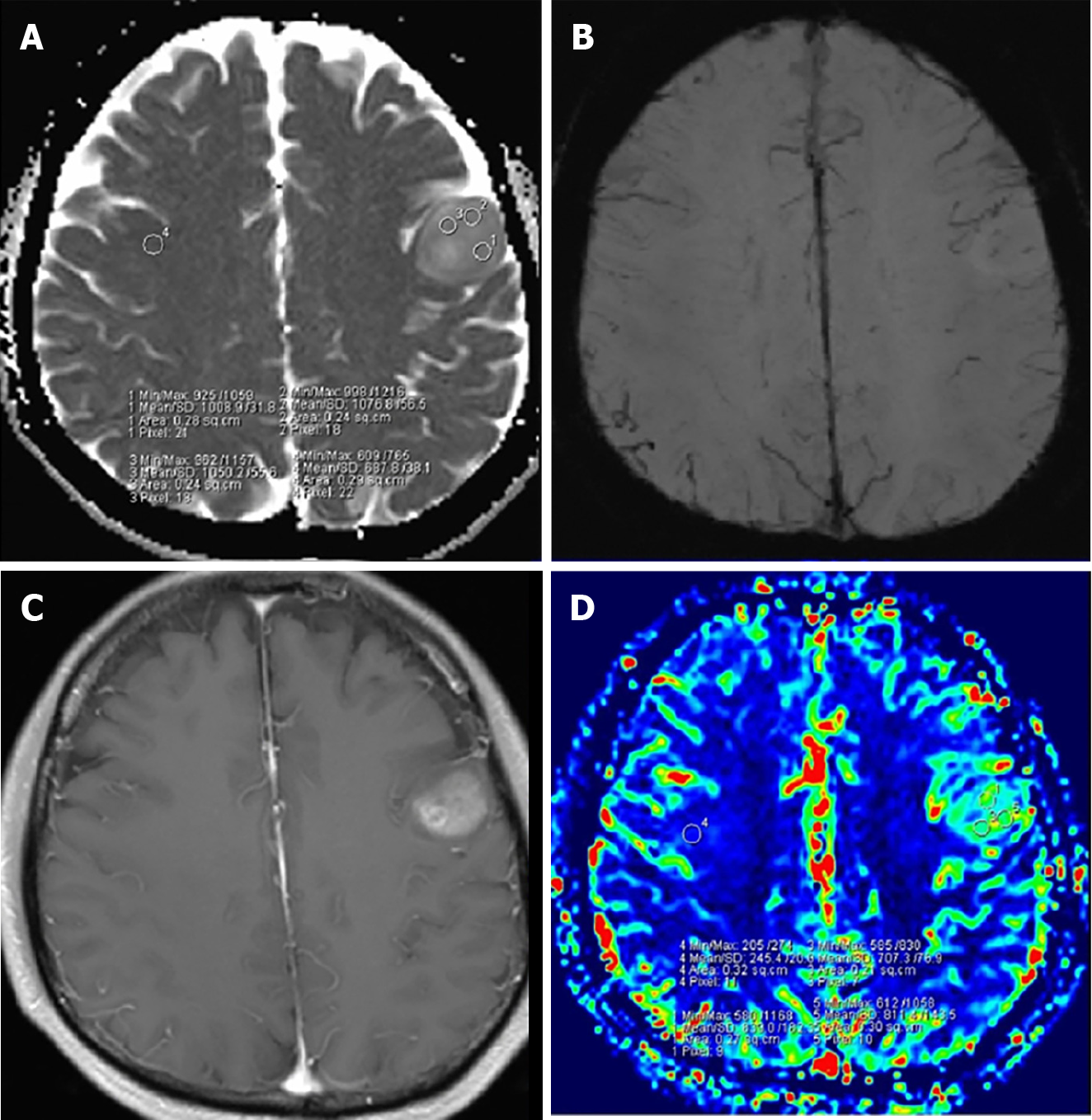
Figure 3 A 53-year-old woman with grade 3 isocitrate dehydrogenase-mutant astrocytoma.
A: Apparent diffusion coefficient (ADC) map shows an increased ADC value in the lesion (ADCmin = 1.016 × 10⁻³ mm2/s); B: Susceptibility weighted imaging shows obvious ITSS in the left parietal region; C: Post-contrast T1WI demonstrates a nonenhancing lesion; D: relative cerebral blood volume (rCBV) map shows the rCBVmax value of 1.19. Citation: Yang X, Xing Z, She D, Lin Y, Zhang H, Su Y, Cao D. Grading of IDH-mutant astrocytoma using diffusion, susceptibility and perfusion-weighted imaging. BMC Med Imaging 2022; 22: 105. Copyright ©2022 The Authors. Published by BioMed Central Ltd[31] (Supplementary material).
- Citation: Mohamed AA, Alshaibi R, Faragalla S, Mohamed Y, Lucke-Wold B. Updates on management of gliomas in the molecular age. World J Clin Oncol 2024; 15(2): 178-194
- URL: https://www.wjgnet.com/2218-4333/full/v15/i2/178.htm
- DOI: https://dx.doi.org/10.5306/wjco.v15.i2.178


