Copyright
©The Author(s) 2024.
World J Clin Oncol. Feb 24, 2024; 15(2): 178-194
Published online Feb 24, 2024. doi: 10.5306/wjco.v15.i2.178
Published online Feb 24, 2024. doi: 10.5306/wjco.v15.i2.178
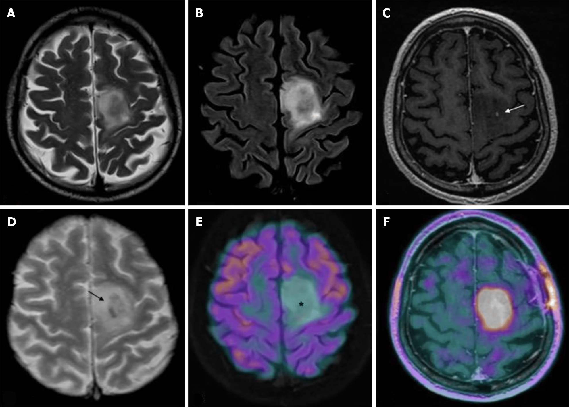
Figure 2 Magnetic resonance imaging and positron emission tomography characteristics of a diffuse isocitrate dehydrogenase mutated 1p/19q codeleted glioma (oligodendroglioma grade 2).
A-F: T2w fast spin echo (A), T2w FLAIR (B), T1w fast spin echo contrast-enhanced (C), T2*-weighted gradient echo (D), 18F-fluorodeoxyglucose (FDG)-positron emission tomography (PET)/magnetic resonance imaging (MRI) (E), and dihydroxyphenylalanine (DOPA)-PET/MRI (F) images are shown; C-F: Mesial frontal mass with a low mass effect and without any significant enhancements after contrast (arrow in C indicates the only spot of blood–brain barrier leakage), and some intratumoral calcifications (arrow in D; computed tomography not shown) with no significant radiotracer uptake by FDG-PET [asterisk in E, but showing a lively uptake of the PET-DOPA radiotracer (F)]. Citation: Feraco P, Franciosi R, Picori L, Scalorbi F, Gagliardo C. Conventional MRI-Derived Biomarkers of Adult-Type Diffuse Glioma Molecular Subtypes: A Comprehensive Review. Biomedicines 2022; 10: 2490. Copyright ©2022 The Authors. Published by MDPI (Basel, Switzerland)[28] (Supplementary material).
- Citation: Mohamed AA, Alshaibi R, Faragalla S, Mohamed Y, Lucke-Wold B. Updates on management of gliomas in the molecular age. World J Clin Oncol 2024; 15(2): 178-194
- URL: https://www.wjgnet.com/2218-4333/full/v15/i2/178.htm
- DOI: https://dx.doi.org/10.5306/wjco.v15.i2.178


