Copyright
©The Author(s) 2020.
World J Clin Oncol. Jul 24, 2020; 11(7): 412-427
Published online Jul 24, 2020. doi: 10.5306/wjco.v11.i7.412
Published online Jul 24, 2020. doi: 10.5306/wjco.v11.i7.412
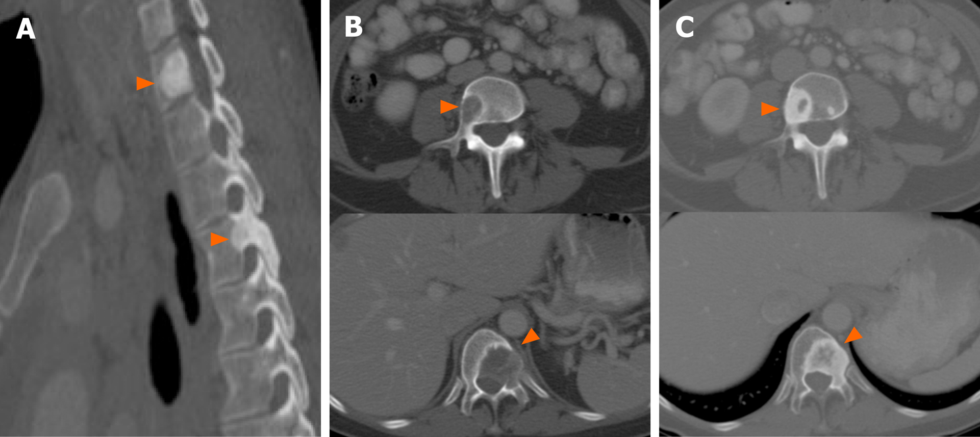
Figure 7 Patterns of bone metastases in non-small cell lung cancer with driver mutations.
A: Pretreatment computed tomography (CT) images (sagittal, bone window) show sclerotic lesions involving the cervicothoracic spine (arrowheads) consistent with osseous metastases in ROS1-positive non-small cell lung cancer (NSCLC); B: Pretreatment CT images (sagittal, bone window) show lytic lesions involving thoracic and lumbar vertebral bodies (arrowheads) in patient with EGFR-mutant NSCLC; C: Post-treatment CT of the same patient as Figure 7B shows interval sclerosis of previously lytic osseous metastases (arrowheads). In general, most NSCLC tend to have lytic lesions in contrast to those with ROS1 or ALK positive NSCLC, which tend to be more sclerotic at presentation. Sclerosis of previously lytic lesion is often seen after treatment.
- Citation: Mendoza DP, Piotrowska Z, Lennerz JK, Digumarthy SR. Role of imaging biomarkers in mutation-driven non-small cell lung cancer. World J Clin Oncol 2020; 11(7): 412-427
- URL: https://www.wjgnet.com/2218-4333/full/v11/i7/412.htm
- DOI: https://dx.doi.org/10.5306/wjco.v11.i7.412


