Copyright
©2012 Baishideng Publishing Group Co.
World J Gastrointest Pathophysiol. Apr 15, 2012; 3(2): 51-59
Published online Apr 15, 2012. doi: 10.4291/wjgp.v3.i2.51
Published online Apr 15, 2012. doi: 10.4291/wjgp.v3.i2.51
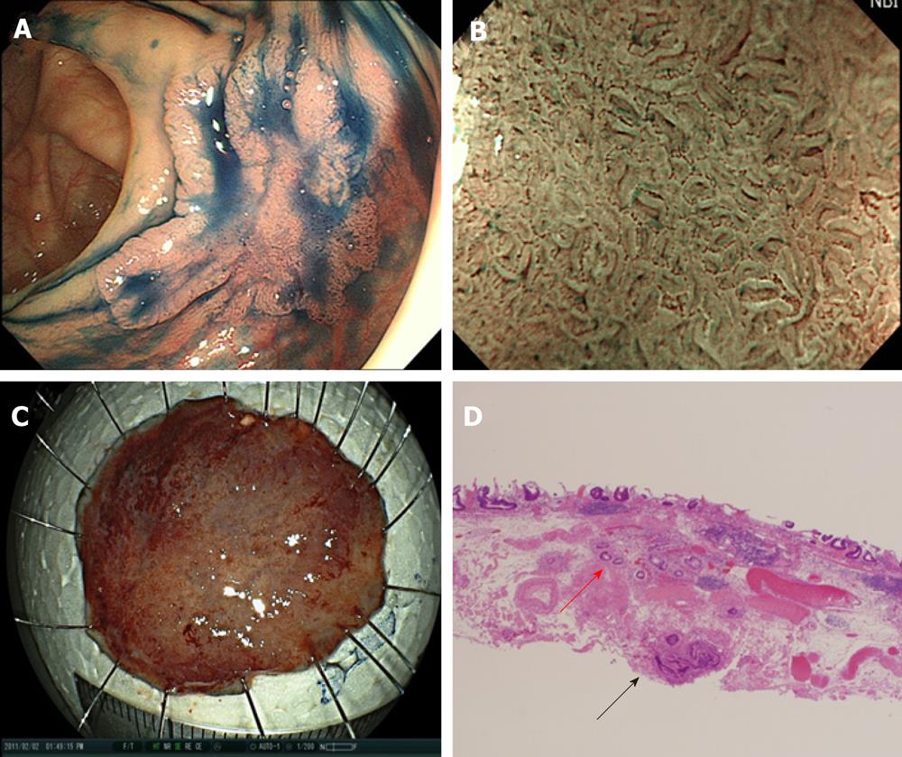
Figure 4 A submucosally invasive cancer with venous infiltration.
A: A tumor graded 0-IIa, measuring 20 mm, located in the ascending colon. The surface of the tumor was slightly depressed (shown by indigo carmine dye); B: Magnifying endoscopy with NBI revealed Type C1/C2 according to Hiroshima classification[43]. The tumor was diagnosed as shallow submucosally invasive cancer and endoscopic submucosal dissection (ESD) was performed; C: En bloc resection was performed. The ESD operation time was 50 min; D: The histopathological diagnosis of the specimen resected by ESD was massive submucosally invasive cancer. The depth of submucosal invasion was 1300 μm and both a positive vertical margin of the tumor (black arrow) and venous infiltration (red arrow) were detected. The appropriate depth of dissection allowed detection of the positive vertical margin and venous infiltration. Additional surgical intervention was performed and no residual tumor or lymph node metastasis was detected. ESD: Endoscopic submucosal dissection; NBI: Narrow band imaging.
- Citation: Yoshida N, Naito Y, Yagi N, Yanagisawa A. Importance of histological evaluation in endoscopic resection of early colorectal cancer. World J Gastrointest Pathophysiol 2012; 3(2): 51-59
- URL: https://www.wjgnet.com/2150-5330/full/v3/i2/51.htm
- DOI: https://dx.doi.org/10.4291/wjgp.v3.i2.51


