Copyright
©The Author(s) 2020.
World J Gastrointest Pathophysiol. May 12, 2020; 11(3): 43-56
Published online May 12, 2020. doi: 10.4291/wjgp.v11.i3.43
Published online May 12, 2020. doi: 10.4291/wjgp.v11.i3.43
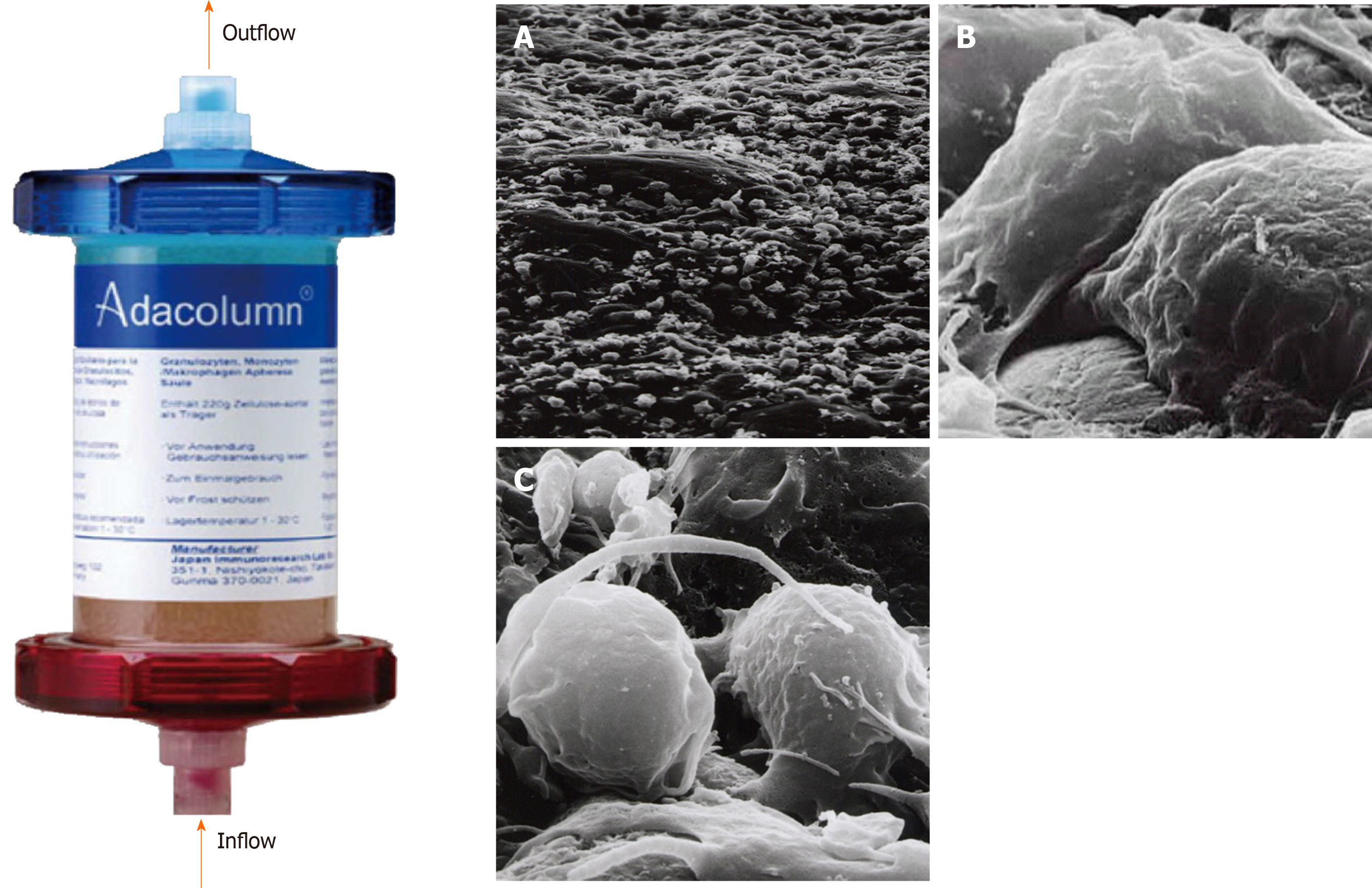
Figure 1 Photograph of Adacolumn and scanning electron photomicrograph of the acetate beads after treatment.
Adacolumn is filled with cellulose acetate beads of 2 mm in diameter (adsorptive carriers) bathed in sterile saline. The blood from the antecubital vein of one arm flows into the column and returns to the antecubital vein in the contralateral arm. A: A low power view (400 ×) of the acetate beads in a column after treatment with cells covering the surface of the carrier; B: Viewed at 10000 ×. Neutrophils were adsorbed onto the beads; C: Viewed at 12000 ×. Activated monocyte/macrophages are seen (taken by Dr. A. Saniabadi of Japan Immunoresearch Laboratories). Modified from reference[31].
- Citation: Chen XL, Mao JW, Wang YD. Selective granulocyte and monocyte apheresis in inflammatory bowel disease: Its past, present and future. World J Gastrointest Pathophysiol 2020; 11(3): 43-56
- URL: https://www.wjgnet.com/2150-5330/full/v11/i3/43.htm
- DOI: https://dx.doi.org/10.4291/wjgp.v11.i3.43


