Copyright
©The Author(s) 2016.
World J Radiol. Sep 28, 2016; 8(9): 799-808
Published online Sep 28, 2016. doi: 10.4329/wjr.v8.i9.799
Published online Sep 28, 2016. doi: 10.4329/wjr.v8.i9.799
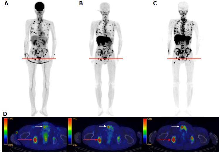
Figure 9 4’-[methyl-11C]-thiothymidine images in multiple myeloma.
18F-FDG (A), 11C-MET (B), and 11C-4DST (C) PET images obtained in a 63-year-old man with multiple myeloma. Numerous active lesions are visible in the three maximum intensity projection images. The fusion images are for the cross-section at the level indicated by the red lines (D). The lesion in the right ischium (red arrow) was positive in all three PET scans. However, the lesion in the right pubis (white arrow) was only positive on the 11C-MET and 11C-4DST PET scans and was equivocal on the 18F-FDG PET scan. Reprinted from Okasaki et al[24], with the permission of Springer. 11C-4DST: 4’-[methyl-11C]-thiothymidine; 11C-MET: 11C-Methionine; 18F-FDG: 2-Deoxy-2-[18F]fluoro-D-glucose; PET: Positron emission tomography.
- Citation: Toyohara J. Evaluation of DNA synthesis with carbon-11-labeled 4′-thiothymidine. World J Radiol 2016; 8(9): 799-808
- URL: https://www.wjgnet.com/1949-8470/full/v8/i9/799.htm
- DOI: https://dx.doi.org/10.4329/wjr.v8.i9.799


