Copyright
©The Author(s) 2016.
World J Radiol. Sep 28, 2016; 8(9): 799-808
Published online Sep 28, 2016. doi: 10.4329/wjr.v8.i9.799
Published online Sep 28, 2016. doi: 10.4329/wjr.v8.i9.799
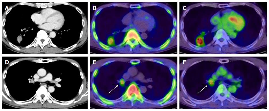
Figure 8 4’-[methyl-11C]-thiothymidine images in non-small cell lung cancer.
Axial images of CT (A), 11C-4DST PET (B), and 18F-FDG PET (C) in a 58-year-old man with lung adenocarcinoma in the right lower lobe. Radioactivity of 11C-4DST in the ascending aorta (representing blood pool) is lower than that of 18F-FDG. Both 11C-4DST and 18F-FDG clearly visualize lung lesions. Right hilar lymph node metastasis is confirmed on CT (D), and uptake of both 11C-4DST PET (E) and 18F-FDG PET (F) identifies the lesion (arrow). However, 11C-4DST images are clearer than 18F-FDG images because of low physiologic 11C-4DST in the mediastinum (blood pool). This research was originally published in JNM[22]. 11C-4DST: 4’-[methyl-11C]-thiothymidine; PET: Positron emission tomography; 18F-FDG: 2-Deoxy-2-[18F]fluoro-D-glucose; CT: Computed tomography.
- Citation: Toyohara J. Evaluation of DNA synthesis with carbon-11-labeled 4′-thiothymidine. World J Radiol 2016; 8(9): 799-808
- URL: https://www.wjgnet.com/1949-8470/full/v8/i9/799.htm
- DOI: https://dx.doi.org/10.4329/wjr.v8.i9.799


