Copyright
©The Author(s) 2016.
World J Radiol. Jun 28, 2016; 8(6): 635-655
Published online Jun 28, 2016. doi: 10.4329/wjr.v8.i6.635
Published online Jun 28, 2016. doi: 10.4329/wjr.v8.i6.635
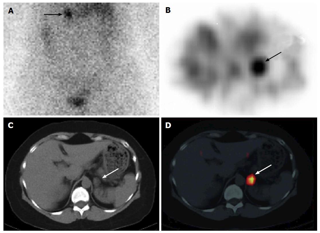
Figure 5 A 55-year-old woman with Cushing syndrome underwent 131I-NP-59 single photon emission computed tomography/computed tomography scan for localization of Cushing’s adenoma.
Planar posterior (A) abdominal image on day 5 post injection of 131I-NP-59 SPECT/CT demonstrates focal uptake in the left adrenal region (arrow). Transverse SPECT (B), CT images (C) and fused SPECT/CT (D) localize activity to a 1.5 cm × 1.8 cm nodule arising from the left adrenal gland (arrows). Findings are compatible with a hyperfunctional left adrenal adenoma. Imaging courtesy of “Adrenal Cortical Imaging with I-131 NP-59 SPECT-CT”. (By Wong et al[181], with permission). SPECT: Single photon emission computed tomography; CT: Computed tomography.
- Citation: Wong KK, Gandhi A, Viglianti BL, Fig LM, Rubello D, Gross MD. Endocrine radionuclide scintigraphy with fusion single photon emission computed tomography/computed tomography. World J Radiol 2016; 8(6): 635-655
- URL: https://www.wjgnet.com/1949-8470/full/v8/i6/635.htm
- DOI: https://dx.doi.org/10.4329/wjr.v8.i6.635


