Copyright
©The Author(s) 2016.
World J Radiol. Jun 28, 2016; 8(6): 581-587
Published online Jun 28, 2016. doi: 10.4329/wjr.v8.i6.581
Published online Jun 28, 2016. doi: 10.4329/wjr.v8.i6.581
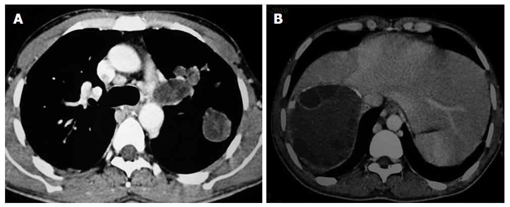
Figure 4 Hydatid cyst on computed tomography.
Axial contrast enhanced computed tomography showing multiple hydatid cysts in left lung (A) and liver (B) (same patient as in Figure 1B). Note peripheral calcification and daughter cysts in liver cyst.
- Citation: Garg MK, Sharma M, Gulati A, Gorsi U, Aggarwal AN, Agarwal R, Khandelwal N. Imaging in pulmonary hydatid cysts. World J Radiol 2016; 8(6): 581-587
- URL: https://www.wjgnet.com/1949-8470/full/v8/i6/581.htm
- DOI: https://dx.doi.org/10.4329/wjr.v8.i6.581


