Copyright
©The Author(s) 2015.
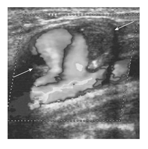
Figure 6 Color Doppler sonogram showing the blood flow of the right common carotid artery, and the haematoma with the rotatory flow within its cavity (arrows).
Note the large neck connecting the carotid to the pseudoaneurysm[90].
- Citation: Mahmoud MZ, Al-Saadi M, Abuderman A, Alzimami KS, Alkhorayef M, Almagli B, Sulieman A. "To-and-fro" waveform in the diagnosis of arterial pseudoaneurysms. World J Radiol 2015; 7(5): 89-99
- URL: https://www.wjgnet.com/1949-8470/full/v7/i5/89.htm
- DOI: https://dx.doi.org/10.4329/wjr.v7.i5.89


