Copyright
©The Author(s) 2015.
World J Radiol. Dec 28, 2015; 7(12): 438-447
Published online Dec 28, 2015. doi: 10.4329/wjr.v7.i12.438
Published online Dec 28, 2015. doi: 10.4329/wjr.v7.i12.438
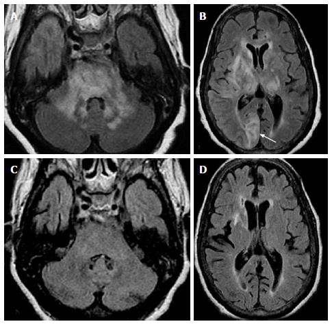
Figure 6 Posterior reversible encephalopathy.
A and B: Axial FLAIR images in an hypertense encephalopathic patient show symmetric bilateral areas of abnormal signal in the MCP and pons. Concomitant involvement of supratentorial subcortical white matter in a “vasogenic type pattern” as well as cortical involvement (arrow on B) was noted; C and D: Three months f/u after control of hypertensive crisis show almost complete resolution of abnormal signal in MCP, pons and supratentorial regions to include the basal ganglia/thalami. Given the asymmetric distribution of supratentorial lesions, PML could be included in the differential. However, cortical involvement is not characteristic for PML. This case could be considered “atypical PRES” and proved by resolution of abnormal white matter disease (compare with Figure 3 where no resolution is noted). PRES: Posterior reversible encephalopathy; MCP: Middle cerebellar peduncles; PML: Progressive multifocal leucoencephalopathy.
- Citation: Morales H, Tomsick T. Middle cerebellar peduncles: Magnetic resonance imaging and pathophysiologic correlate. World J Radiol 2015; 7(12): 438-447
- URL: https://www.wjgnet.com/1949-8470/full/v7/i12/438.htm
- DOI: https://dx.doi.org/10.4329/wjr.v7.i12.438


