Copyright
©2014 Baishideng Publishing Group Inc.
World J Radiol. Sep 28, 2014; 6(9): 657-668
Published online Sep 28, 2014. doi: 10.4329/wjr.v6.i9.657
Published online Sep 28, 2014. doi: 10.4329/wjr.v6.i9.657
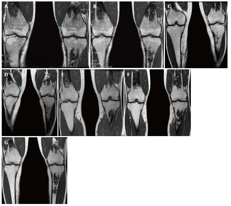
Figure 15 Bone marrow improvement.
Gaucher Type 1 patient with N370S/N370S genotype who started enzyme replacement therapy (ERT) in 1997. Coronal T1 WI of the distal femora and proximal tibiae in 1998 (A), 1999 (B), 2002 (C), 2003 (D), 2006 (E), 2008 (F) and 2011 (G). Medullary infarcts are partially seen in both distal femurs and the left tibia. The low T1 SI significantly improves between 1998 and 2002 with near normal marrow signal seen in the noninfarcted areas in 2008 and years later. Although this patient has the same genotype as the patient in Figure 14, there is a longer time to improvement indicating the limited genotype/phenotype correlation.
- Citation: Simpson WL, Hermann G, Balwani M. Imaging of Gaucher disease. World J Radiol 2014; 6(9): 657-668
- URL: https://www.wjgnet.com/1949-8470/full/v6/i9/657.htm
- DOI: https://dx.doi.org/10.4329/wjr.v6.i9.657


