Copyright
©2014 Baishideng Publishing Group Inc.
World J Radiol. Sep 28, 2014; 6(9): 636-642
Published online Sep 28, 2014. doi: 10.4329/wjr.v6.i9.636
Published online Sep 28, 2014. doi: 10.4329/wjr.v6.i9.636
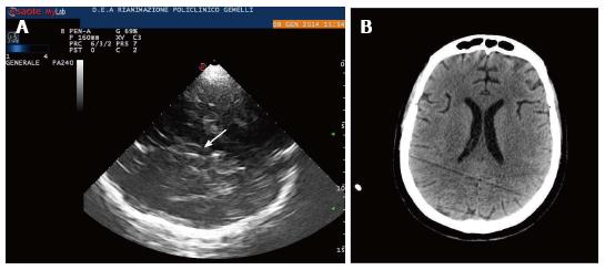
Figure 4 Lateral ventricles.
A: Lateral ventricles in echography. Frontal horns of lateral ventricles are visualized as hypoechogenic structure, well visible between two parallel lines corresponding to the medial and lateral layer of the ependima. The three parallel lines correspond to lateral layers of ependima and septum pellucidum. The image is generated by a sectorial probe, and lateral ventricle ipsilateral to the probe is depicted at the same depth of the third ventricle. Arrow shows third ventricle, Small arrow heads on the left show frontal horns of lateral ventricles; B: Lateral ventricles in computed tomography (CT). CT scan of lateral ventricles correspondent of Figure 4A is shown.
- Citation: Caricato A, Pitoni S, Montini L, Bocci MG, Annetta P, Antonelli M. Echography in brain imaging in intensive care unit: State of the art. World J Radiol 2014; 6(9): 636-642
- URL: https://www.wjgnet.com/1949-8470/full/v6/i9/636.htm
- DOI: https://dx.doi.org/10.4329/wjr.v6.i9.636


