Copyright
©2014 Baishideng Publishing Group Inc.
World J Radiol. Jul 28, 2014; 6(7): 446-458
Published online Jul 28, 2014. doi: 10.4329/wjr.v6.i7.446
Published online Jul 28, 2014. doi: 10.4329/wjr.v6.i7.446
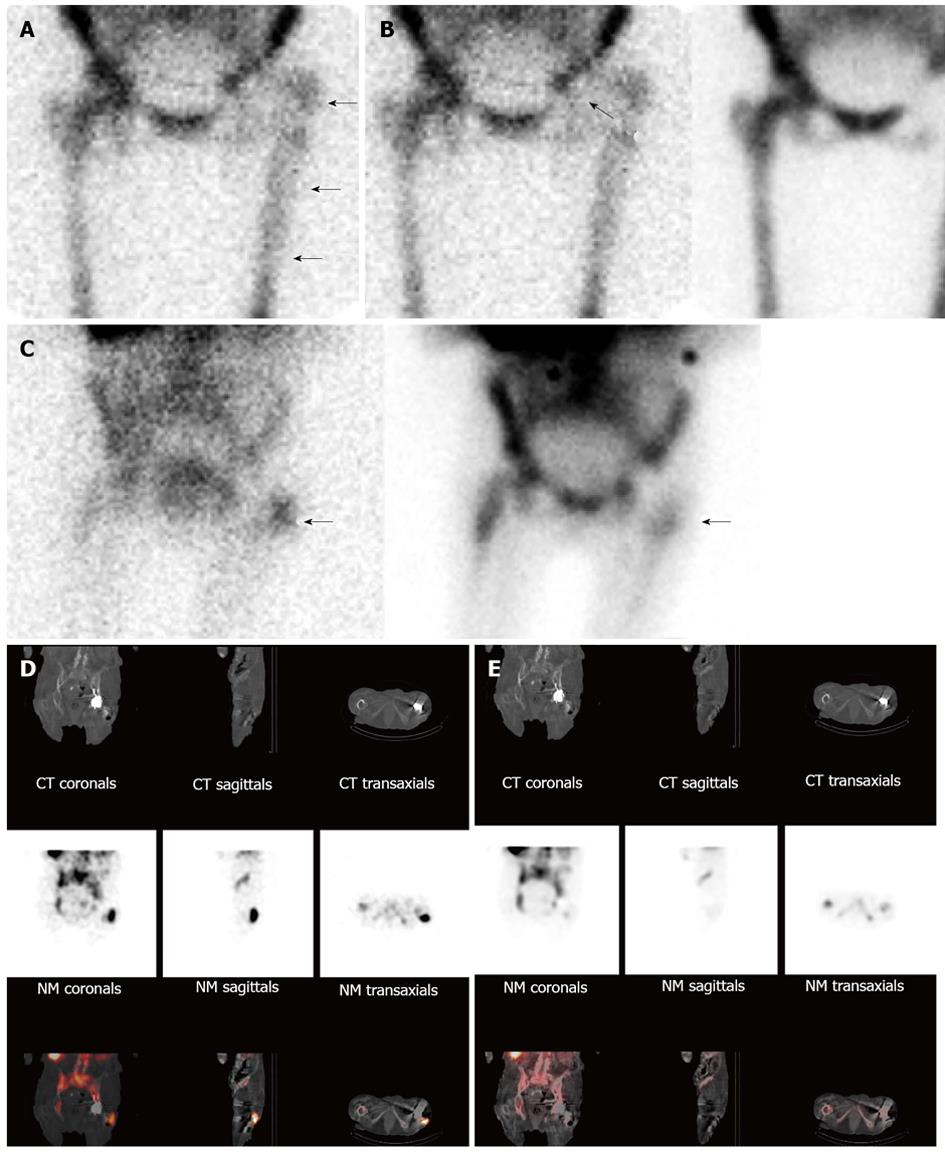
Figure 7 Infected left hip arthroplasty.
A: On this anterior image from an indium-111 labeled leukocyte study, periprosthetic activity (arrows) is similar in intensity to activity in the contralateral lower extremity and less intense than pelvic activity, areas typically used as reference points when interpreting these studies. Because studies in which the intensity of labeled leukocyte activity in the region of interest does not exceed intensity of activity in the reference point, this study could be erroneously interpreted as negative for infection; B: The distribution of periprosthetic activity on the labeled leukocyte (left, 111In-WBC) and sulfur colloid bone marrow (right, 99mTc-SC) images is spatially incongruent (arrows), i.e., there is activity in the left hip joint on the labeled leukocyte image, but not on the bone marrow image. The combined study is positive for infection. (Same patient illustrated in Figure 7A); Although the planar combined indium labeled leukocyte/bone marrow study (C, left, 111In-WBC; right, 99mTc-SC) is positive for infection (arrows), precise information about the location and extent of infection is lacking. On the fused images (bottom row) from the labeled leukocyte SPECT/CT (D) the location of the abnormal labeled leukocyte accumulation (arrows) can clearly be seen adjacent and extending to the prosthesis at the level of the greater trochanter. Note also the adjacent hypodense area in the soft tissues, consistent with abscess. Bone marrow SPECT/CT images (E) acquired simultaneously with the labeled leukocyte images in 16a confirm that the activity on the labeled leukocyte component of the examination is due to infection. Whether or not the bone marrow component of the SPECT/CT study contributes additional information beyond what planar imaging provides remains to be determined.
- Citation: Palestro CJ. Nuclear medicine and the failed joint replacement: Past, present, and future. World J Radiol 2014; 6(7): 446-458
- URL: https://www.wjgnet.com/1949-8470/full/v6/i7/446.htm
- DOI: https://dx.doi.org/10.4329/wjr.v6.i7.446


