Copyright
©2014 Baishideng Publishing Group Inc.
World J Radiol. Jul 28, 2014; 6(7): 446-458
Published online Jul 28, 2014. doi: 10.4329/wjr.v6.i7.446
Published online Jul 28, 2014. doi: 10.4329/wjr.v6.i7.446
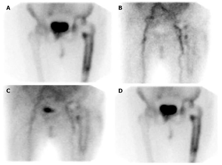
Figure 3 Aseptically loosened left hip replacement.
A: On the 99mTc-MDP bone scan, there is diffusely increased radiopharmaceutical accumulation around the femoral component of the cemented 2 years old prosthesis. Compare with Figure 2A; B-D: On the 99mTc-MDP bone scan, there is diffuse hyperperfusion, and hyperemia around the prosthesis on the flow and blood pool images, and diffusely increased periprosthetic radiopharmaceutical on the delayed, bone image (same patient illustrated in Figure 3A), B: Flow; C: Blood pool; D: Delayed. The scan appearance is nearly identical to that of the infected prosthesis in Figure 2B.
- Citation: Palestro CJ. Nuclear medicine and the failed joint replacement: Past, present, and future. World J Radiol 2014; 6(7): 446-458
- URL: https://www.wjgnet.com/1949-8470/full/v6/i7/446.htm
- DOI: https://dx.doi.org/10.4329/wjr.v6.i7.446


