Copyright
©2014 Baishideng Publishing Group Inc.
World J Radiol. Jul 28, 2014; 6(7): 446-458
Published online Jul 28, 2014. doi: 10.4329/wjr.v6.i7.446
Published online Jul 28, 2014. doi: 10.4329/wjr.v6.i7.446
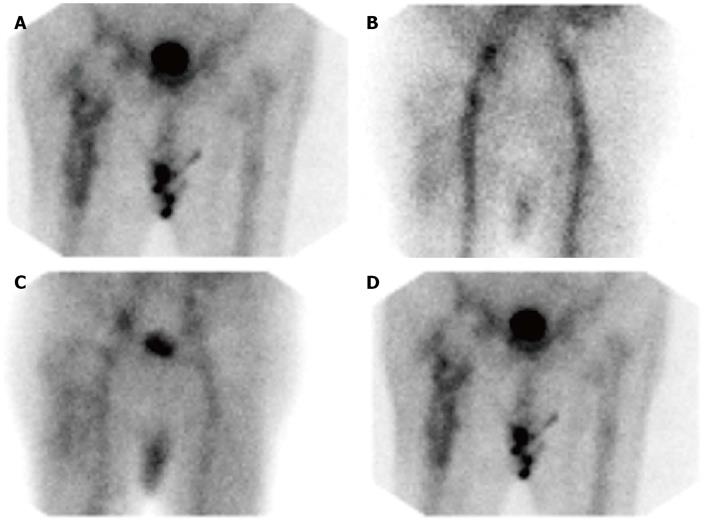
Figure 2 Infected right hip arthroplasty.
A: On the 99mTc-methylene diphosphonate bone scan, there is irregularly increased radiopharmaceutical accumulation around the entire femoral component of the 2 years old cementless (revision) prosthesis, a pattern which some investigators have reported as specific for infection; B-D: On the 99mTc-MDP bone scan, there is diffuse hyperperfusion, and hyperemia around the prosthesis on the flow and blood pool images, and diffusely increased periprosthetic radiopharmaceutical on the delayed, bone image (same patient illustrated in Figure 2A); B: Flow; C: Blood pool; D: Bone.
- Citation: Palestro CJ. Nuclear medicine and the failed joint replacement: Past, present, and future. World J Radiol 2014; 6(7): 446-458
- URL: https://www.wjgnet.com/1949-8470/full/v6/i7/446.htm
- DOI: https://dx.doi.org/10.4329/wjr.v6.i7.446


