Copyright
©2014 Baishideng Publishing Group Inc.
World J Radiol. Oct 28, 2014; 6(10): 779-793
Published online Oct 28, 2014. doi: 10.4329/wjr.v6.i10.779
Published online Oct 28, 2014. doi: 10.4329/wjr.v6.i10.779
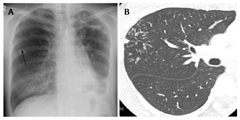
Figure 6 Postprimary tuberculosis in a woman in her 40s.
A: Chest radiograph shows faint nodular opacities in the right middle lung field (arrow). There is also volume loss of the left lung with patchy consolidations and thickening of the pleura and possible left pleural effusion, indicative of old tuberculosis; B: Thin-section computed tomography demonstrates centrilobular branching opacities (tree-in-bud appearance) in the right upper lobe (arrows). The branching opacities are denser, more distributed and more peripherally located than those of ordinary bronchopneumonia (compare with Figure 3).
- Citation: Nambu A, Ozawa K, Kobayashi N, Tago M. Imaging of community-acquired pneumonia: Roles of imaging examinations, imaging diagnosis of specific pathogens and discrimination from noninfectious diseases. World J Radiol 2014; 6(10): 779-793
- URL: https://www.wjgnet.com/1949-8470/full/v6/i10/779.htm
- DOI: https://dx.doi.org/10.4329/wjr.v6.i10.779


