Copyright
©2013 Baishideng Publishing Group Co.
World J Radiol. Oct 28, 2013; 5(10): 356-371
Published online Oct 28, 2013. doi: 10.4329/wjr.v5.i10.356
Published online Oct 28, 2013. doi: 10.4329/wjr.v5.i10.356
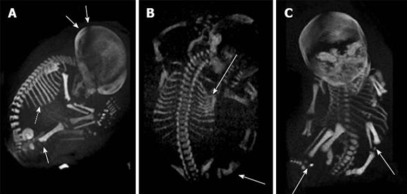
Figure 8 Prenatal diagnosis by three-dimensional helical computer tomography.
A: Achondroplasia (sagittal view): macrocephaly (double arrows), short ribs (dotted arrow) and increased thickness of the femoral metaphysis (single arrow); B: Osteogenesis imperfecta (posterior view): fractures of ribs and femur (arrows); C: Chondrodysplasia punctata (frontal view): epiphyseal calcifications of long bones (arrows). Adapted from Ruano et al[102].
- Citation: Renna MD, Pisani P, Conversano F, Perrone E, Casciaro E, Renzo GCD, Paola MD, Perrone A, Casciaro S. Sonographic markers for early diagnosis of fetal malformations. World J Radiol 2013; 5(10): 356-371
- URL: https://www.wjgnet.com/1949-8470/full/v5/i10/356.htm
- DOI: https://dx.doi.org/10.4329/wjr.v5.i10.356


