Copyright
©2013 Baishideng Publishing Group Co.
World J Radiol. Oct 28, 2013; 5(10): 356-371
Published online Oct 28, 2013. doi: 10.4329/wjr.v5.i10.356
Published online Oct 28, 2013. doi: 10.4329/wjr.v5.i10.356
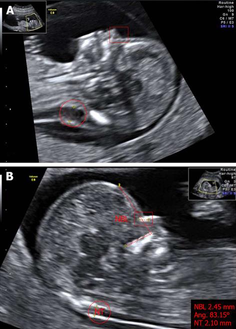
Figure 2 Markers of chromosomal defects.
A: Fetus with Down’s syndrome: increased NT (red circle), and absent nasal bone (red square where nasal bone was expected) at 11 wk of pregnancy; B: Normal fetus: measurements of nuchal translucency (NT, red circle), facial angle (red dashed line) and nasal bone length (NBL, red square) at 13 wk of pregnancy. The image has been certified by the Fetal Medicine Foundation. Photos taken by Wolfgang Moroder. Creative Commons[113].
- Citation: Renna MD, Pisani P, Conversano F, Perrone E, Casciaro E, Renzo GCD, Paola MD, Perrone A, Casciaro S. Sonographic markers for early diagnosis of fetal malformations. World J Radiol 2013; 5(10): 356-371
- URL: https://www.wjgnet.com/1949-8470/full/v5/i10/356.htm
- DOI: https://dx.doi.org/10.4329/wjr.v5.i10.356


