Copyright
©2012 Baishideng Publishing Group Co.
World J Radiol. Aug 28, 2012; 4(8): 353-371
Published online Aug 28, 2012. doi: 10.4329/wjr.v4.i8.353
Published online Aug 28, 2012. doi: 10.4329/wjr.v4.i8.353
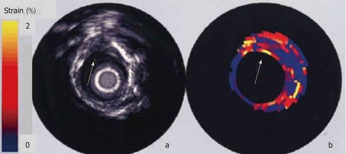
Figure 9 In vitro intravascular echogram and elastogram of a human femoral artery.
The elastogram reveals that the plaque at 12 o’clock contains a soft core that is covered from the lumen by a stiff cap. At 9 o’clock a soft tissue is present at the lumen vessel-wall boundary. A different strain was found at 9 and 3 o’clock and this difference was not present in the echogram[140].
- Citation: Soloperto G, Casciaro S. Progress in atherosclerotic plaque imaging. World J Radiol 2012; 4(8): 353-371
- URL: https://www.wjgnet.com/1949-8470/full/v4/i8/353.htm
- DOI: https://dx.doi.org/10.4329/wjr.v4.i8.353


