Copyright
©2012 Baishideng Publishing Group Co.
World J Radiol. Aug 28, 2012; 4(8): 353-371
Published online Aug 28, 2012. doi: 10.4329/wjr.v4.i8.353
Published online Aug 28, 2012. doi: 10.4329/wjr.v4.i8.353
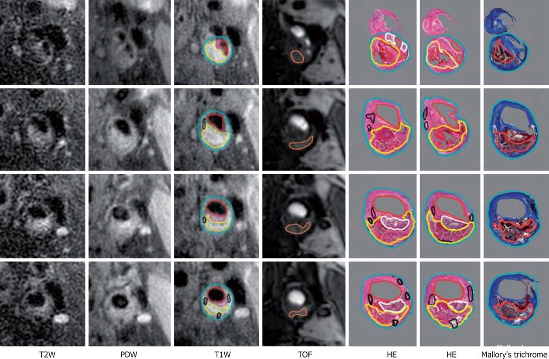
Figure 4 Example of histological validation of magnetic resonance imaging at four consecutive locations spanning the bifurcation.
Multiple histological sections (at 0.5-1.0 mm-separation) generally correspond to each 2-mm thick image. Contours have been drawn for lumen (red), outer wall (cyan), lipid-rich/necrotic core (yellow), calcification (black), loose fibrous matrix (pink/white) and hemorrhage (orange)[49]. TOF: Time-of flight; HE: Hematoxylin and eosin; PDW: Proton density weighted; T2W: T2-weighted; T1W: T1-weighted.
- Citation: Soloperto G, Casciaro S. Progress in atherosclerotic plaque imaging. World J Radiol 2012; 4(8): 353-371
- URL: https://www.wjgnet.com/1949-8470/full/v4/i8/353.htm
- DOI: https://dx.doi.org/10.4329/wjr.v4.i8.353


