Copyright
©2010 Baishideng Publishing Group Co.
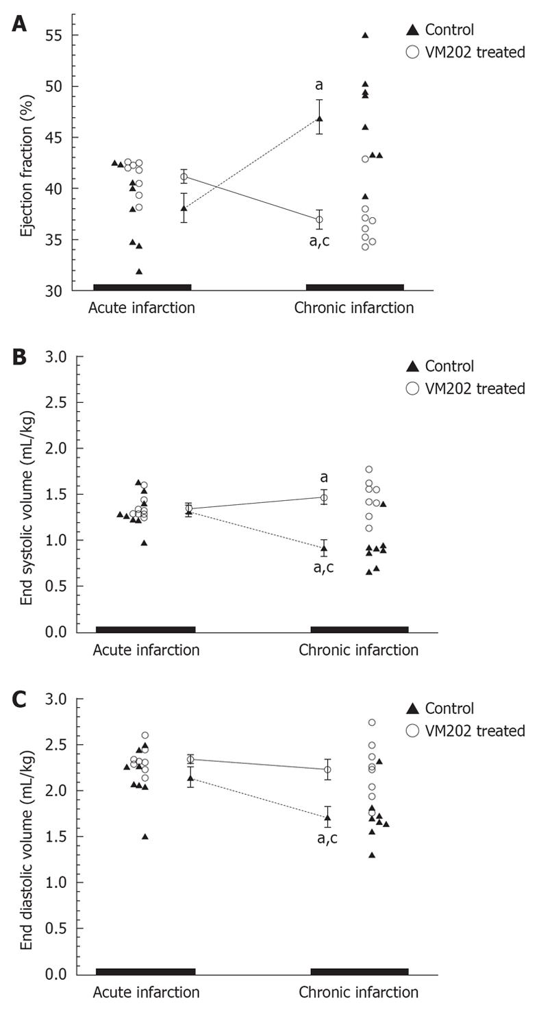
Figure 3 The ejection fraction (A), end systolic volumes (B) and end diastolic volumes (C) are shown for control and hepatocyte growth factor gene (VM202) treated animals.
The hepatocyte growth factor gene administered at 3 d after reperfusion significantly decreased end diastolic (mL/kg) and end systolic volumes at 8 wk compared to 3 d infarct (aP < 0.05) and control group (cP < 0.05). Control animals at 8 wk showed a significant decrease in ejection fraction and significant increase in end systolic and end diastolic volumes compared with 3 d infarct[59].
- Citation: Saeed M, Hetts S, Wilson M. Reperfusion injury components and manifestations determined by cardiovascular MR and MDCT imaging. World J Radiol 2010; 2(1): 1-14
- URL: https://www.wjgnet.com/1949-8470/full/v2/i1/1.htm
- DOI: https://dx.doi.org/10.4329/wjr.v2.i1.1


