Copyright
©The Author(s) 2016.
World J Cardiol. Sep 26, 2016; 8(9): 520-533
Published online Sep 26, 2016. doi: 10.4330/wjc.v8.i9.520
Published online Sep 26, 2016. doi: 10.4330/wjc.v8.i9.520
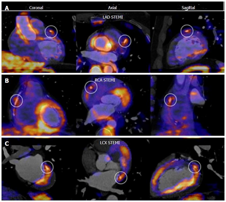
Figure 7 Fluorodeoxyglucose positron emission tomography of the coronary arteries.
PET CT fusion imaging in three cases of patients with STEMI. An increased 18F-FDG uptake at stent site is shown in different culprit vessels, from A to C: LAD, RCA and LCX. Adapted with permission from Cheng et al[107]. This research was originally published in JNM. ©by the Society of Nuclear Medicine and Molecular Imaging, Inc. FDG: Fluorodeoxyglucose; PET: Positron emission tomography; STEMI: ST elevation myocardial infarction; LAD: Left anterior descending coronary artery; RCA: Right coronary artery; LCX: Left circumflex coronary artery.
- Citation: Pozo E, Agudo-Quilez P, Rojas-González A, Alvarado T, Olivera MJ, Jiménez-Borreguero LJ, Alfonso F. Noninvasive diagnosis of vulnerable coronary plaque. World J Cardiol 2016; 8(9): 520-533
- URL: https://www.wjgnet.com/1949-8462/full/v8/i9/520.htm
- DOI: https://dx.doi.org/10.4330/wjc.v8.i9.520


