Copyright
©The Author(s) 2016.
World J Cardiol. Sep 26, 2016; 8(9): 520-533
Published online Sep 26, 2016. doi: 10.4330/wjc.v8.i9.520
Published online Sep 26, 2016. doi: 10.4330/wjc.v8.i9.520
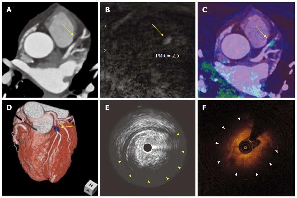
Figure 6 T1 hyperintense coronary plaques in cardiac magnetic resonance.
Noninvasive and invasive coronary imaging of a significant plaque in proximal LAD. CCTA (A) showed a noncalcified plaque in LAD causing significant stenosis. When noncontrast T1-weighted CMR imaging was performed (B) a hyperintense lesion was detected. Afterwards, CMR images were fused with CCTA (C and D) and this lesion was found to correspond with the previously described coronary stenosis. Interestingly, during the subsequent coronary angiography it showed a large lipid component in IVUS (E) as well as OCT (F). Adapted with permission from Asaumi et al[106]. LAD: Left anterior descending coronary artery; CCTA: Coronary computed tomography; CMR: Cardiac magnetic resonance; IVUS: Intravascular ultrasound; OCT: Optical coherence tomography; PMR: Plaque to myocardium signal intensity ratio.
- Citation: Pozo E, Agudo-Quilez P, Rojas-González A, Alvarado T, Olivera MJ, Jiménez-Borreguero LJ, Alfonso F. Noninvasive diagnosis of vulnerable coronary plaque. World J Cardiol 2016; 8(9): 520-533
- URL: https://www.wjgnet.com/1949-8462/full/v8/i9/520.htm
- DOI: https://dx.doi.org/10.4330/wjc.v8.i9.520


