Copyright
©The Author(s) 2016.
World J Cardiol. Nov 26, 2016; 8(11): 623-637
Published online Nov 26, 2016. doi: 10.4330/wjc.v8.i11.623
Published online Nov 26, 2016. doi: 10.4330/wjc.v8.i11.623
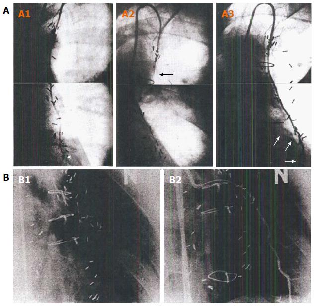
Figure 3 Composite of string sign data.
Angiographic documentation of the development of a “string-sign” IMA graft. A: Data from Dincer et al[98]. A1: Composite still images from angiogram of IMA-LAD graft at 8 d postop. White arrow show anastomotic site; A2: Composite still images from an angiogram at 1-year postop. Black arrow identifies “string-sign” IMA conduit with little if any distal flow; A3: Composite still images from angiogram at 5 years postop, documenting a widely patent IMA-LAD graft. The three white arrows outline the native TVECA LAD proximal and distal to the anastomosis. There is no angiographic evidence of atherosclerotic disease in the IMA, and no anastomotic evidence of narrowing; B: Data from Kitamura et al[99]. Images that clearly illustrate the physiology of arterial conduits. B1: A stringlike LIMA with no-flow into the LAD. Because of the limitations of conventional angiography, flow down the TVECA LAD cannot be simultaneously visualized, but was patent with good antegrade flow; B2: Repeat LIMA arteriography now showing anatomical patency of the graft, as a result of temporary occlusion of the recipient LAD with a percutaneous transluminal coronary angioplasty balloon. The acute influence of anterior wall hypoperfusion immediately translated into resumed functionality of the LIMA graft, documenting the coupling of physiologic IMA flow to the distal regional myocardial physiologic status. IMA: Internal mammary artery; LAD: Left anterior descending coronary artery; TVECA: Target vessel epicardial coronary artery.
- Citation: Ferguson Jr TB. Physiology of in-situ arterial revascularization in coronary artery bypass grafting: Preoperative, intraoperative and postoperative factors and influences. World J Cardiol 2016; 8(11): 623-637
- URL: https://www.wjgnet.com/1949-8462/full/v8/i11/623.htm
- DOI: https://dx.doi.org/10.4330/wjc.v8.i11.623


